User:K huynhh/sandbox
| This is not a Wikipedia article: It is an individual user's work-in-progress page, and may be incomplete and/or unreliable. For guidance on developing this draft, see Wikipedia:So you made a userspace draft. Find sources: Google (books · news · scholar · free images · WP refs) · FENS · JSTOR · TWL |
Introduction
[edit]Zebrafish have proved to be an effective model organism in a number of different fields of studies. Their transparent and externally mass-produced embryos are affordable. Zebrafish are similar to the human genome, due to the fact that they are also vertebrates. They have a short life cycle, reaching maturation at 90 days past fertilization. Fertilization in zebrafish happens after oocyte maturation. Eggs are fertilized externally, which initiates embryonic development. Cleavage begins soon after fertilization, a process of mass cell division. Gastrulation in zebrafish forms the three germ layers; ectoderm, mesoderm, and endoderm. Neurulation is the process of neural tube formation. Axis signaling consists of multiple genes responsible for the formation of the anterior-posterior and dorsal-ventral axes. There are both advantages and disadvantages of the zebrafish model system.
Life Cycle
[edit]
The life cycle of a zebrafish is extremely fast. It normally takes about 3 months for the zebrafish to become an adult. It starts out as an embryo, which all offspring are considered embryos until they are born or hatch. [1] The embryo normally hatches around 48-72 hours after fertilization. The larva is neither an embryo or a juvenile. [1] The juvenile state normally lasts about 4-12 weeks after fertilization, it just depends on the living situations. This state is where the fish has all the adult characteristics, except it is still not sexually mature. [1]The Zebrafish can’t reproduce at this stage. The adult stage is when the fish can breed or reproduce. Metamorphosis is the step where a juvenile is formed into an adult fish. They lose some larval features during this stage. [1] The post-embryonic development is basically any growith that occurs after embryogenesis. You can see in figure 1 all the individual steps of life cycle in the Zebrafish.
Fertilization in Zebrafish
[edit]Oocyte Maturation
Zebrafish must go through processes for oocyte maturation before fertilization can begin. The female oocyte goes through four stages before progressing to full maturation. [2] Stages I, II, and III the oocyte grows, and some maternal mRNA is distributed throughout the oocyte. During oocyte maturation some mRNA is specifically localized for future development of the animal/vegetal axis. [2] There is a difference in the size of the mature oocytes between species. The zebrafish oocytes are anywhere from 7-700 micrometers (0.74 m). Zebrafish oocytes are transcriptionally active, which means active gene transcription takes place. [3] Zebrafish egg form a Balbiani body, which is a visible cloud of mitochondria. The gene bucky ball (buc) is essential for the Balbiani body formation in zebrafish. [4] A study has shown that without buc, the zebrafish egg is unable to form a Balbiani body, which affects the animal-vegetal polarity. [4]
Yolk
The oocyte forms yolk granules for preparation of fertilization. The mature oocyte in zebrafish is released when a rise in gonadotrophin and a steroid is produced. [3] The nucleus resides in the animal pole of the oocyte. Fertilization in zebrafish are external, meaning the eggs and sperm are released outside of the body for fertilization.
Sperm
The zebrafish sperm does not have an acrosome. The sperm enter through the micropyle usually near the animal pole on the oocyte, which allows the sperm to penetrate the chorion. [5] At this point, fertilization has begun. After the sperm enters the oocyte through the micropyle, there is a rise in calcium in the newly fertilized egg. The calcium transientspreads from the sperm entry to the animal pole. [5] A study was done by researchers to show the analysis of calcium signaling in the zebrafish oocyte and when the calcium is released. The rise in calcium is important for the rise of animal pole, after oocyte has been fertilized. In the study, images show the rise in calcium by calcium green fluorescence. At 2.0 min after sperm was injected, the calcium was most apparent. [5] The sperm can only enter through this small hole, known as the micropyle on the female egg. The micropyle allows for only one sperm to enter inside the oocyte. The gene buc prevents polyspermy from happening, by not forming multiple micropyles. [6]
Zygote
Once the male sperm haploid genome is released into the zygote, the gene futile cycle (fue) is required for female nuclear migration toward the sperm nucleus. [7] After the egg is fertilized and the nuclei are fused together forming a diploid zygote, a cytoplasmic cap is formed at the top of the animal pole, in preparation for cleavage. [3]
Cleavage
[edit]
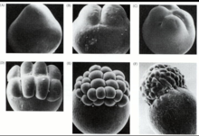
Cleavage in zebrafish happens in the blastodisc. [8] The divisions don’t divide the egg entirely .[8] Cleavage is when a cell divides. The embryo develops the cytoplasm of the blastodisc which is called discoidal. [8] Figure 2 shows the process of cleavage in many different steps. Cleavage happens every 15 minutes. The first 5 divisions are vertical and creates 32 cells. [9] The actin is activated and changes the embryo shape from spherical to pear shaped. The first 12 divisions form a mound of cells in the animal pole in the yolk cell. [8] These cells make up the blastoderm. During the 10th cell division the zygotic gene transcription begins, and you can see three distinct cell populations.
The Yolk syncytial layer (YSL) is found during the 9th or 10th division. There are two parts of the yolk syncytial, which is external and internal. The internal is formed from the nuclei going under the blastoderm. [8]The external is formed in front of the blastoderm. The enveloping layer is the outer cells of the blastoderm. The enveloping layer will later transform into the periderm which is just protection for the cell. The cells in between the enveloping layer and the yolk syncytial layer are called the deep cells. [8] The blastoderm is set on what it will become before gastrulation happens.
Gastrulation
[edit]
Gastrulation is the next top following cleavage. Three cell movements are essential during during gastrulation are: epiboly, involution, and convergent extension.[3] The standard and desired result of gastrulation is the formation of three germ layers: ectoderm, mesoderm, and endoderm.[10] Beginning with epiboly, the cells of the blastoderm thin out and spread over the yolk.[11] The blastoderm cells covering the yolk in the animal pole is termed the internal yolk syncytial layer, or internal YSL.[11] The yolk cell in the vegetal pole where blastoderm cells have not enveloped is termed the external yolk syncytial layer, or external YSL.[11] Epiboly of cells continue and eventually cover the animal hemisphere of the cell.[3] Stages of epiboly are marked as percentages. Once epiboly has reached 50%, typically 5-6 hours[3], involution begins and the mesendodermal cell layer forms, also called the hypoblast.[11] The second cell movement is involution of cells.[3] Deep cells start to move inward near the blastoderm of the cell.[3] This involution creates the epiblast and hypoblast.[3] As involution continues, on the dorsal side of the embryo, it begins to thicken and forms the embryonic shield. This structure can also be compared to the dorsal lip, as seen in other organisms.[3] As cells continue to involute, the endoderm and mesoderm form. Simultaneously, on the dorsal side of the embryo, during convergent extension of the embryonic shield, cells narrow and elongate perpendicularly.[10] The midline of dorsal region elongates to become of the midline of the anteroposterior axis. The midventral sector forms the tailbud instead of cells participating in convergent extension.[3] The Wnt-PCP pathway is an important gene expression pathway for the convergent extension of cells. Epiboly and involution take about 10 hours to complete.[3]
Neurulation in Zebrafish
[edit]Neurulation is the development of a neural tube from the ectoderm layer, which is stated in gastrulation, and the neural plate. [12]
After gastrulation, the mesodermal layer meets the ectoderm, inducing neural induction. Neural induction is the first step in the neural tube formation. [13]
Neural plate
Some signals that are expressed early in neurogenesis is Wnt, Fgf , Nodal proteins, and retinoic acid. These are pattern signals that help with axis formation during the formation of the neural plate. [13] A prechordal plate and the lateral region form from the central part of embryo. The dorsal ectoderm later becomes the neural plate. [3] The neural plate is a flat sheet of neuroepithelium cells, which comes from the thickening of epiblast cells. [14] The lateral ends of the neural plate begin to thicken, and fuse together forming a neural keel. [15]
Neural tube
The neural keel then fuses at the midline, forming the neural rod. This process happens at about 16 hours of embryo development, forming a solid neural rod from the neural plate. There are some structural proteins, Pard3 and Rab11a involved in the midline polarization, during the formation of the neural rod. [13] The neural rod goes through a process called secondary neurulation to form the neural tube. During neurogenesis, the mesoderm contains somites that are continuously differentiating for future organ development. [3]
Early Development Leading to Axis Formation
[edit]
At ten hours post fertilization, the zebrafish embryo has clearly recognizable anterior-posterior and dorsal-ventral axis [16]. To generate this basic body plan, the zebrafish, the embryo undergoes rapid development and morphogenetic changes [16]. A blastodisc is then formed on top of the yolk after fertilization, during the following three hours of development, rapid, synchronous cleavage occurs within the blastodisc to generate a blastula embryo of around 1000 cells [17]. After the mid blastula transition, at about four hours post fertilization (hpf), cell rearrangements reshape the blastoderm into what will be a characteristic vertebrate body plan. In the process known as epiboly, cells interpolate radially, thinning the blastoderm over the yolk. After gastrulation, epiboly movements have spread the blastomeres so that the blastoderm covers the entire yolk cell. There are also three other movements that contribute to the formation of the axis. At five hours post fertilization, cells at the margin internalize and form the hypoblast, the precursor of the mesoderm and endoderm [18].
By six hpf, movements of convergence and extension have begun, which results in dorsal accumulation of cells moving from lateral and ventral regions of the blastoderm (convergence) [19]. Converging cells then insert with dorsal blastomeres and spread them along the animal-vegetal aces, which leads to the lengthening of the anterior-posterior axis (extension). This convergence of all these cells to the dorsal side leads to the first clearly apparent break in the radial symmetry and forms the shield, a thickening at the dorsal blastoderm margin that is equivalent of the amphibian Spemann-Mangold organizer [19].
Advantages and Disadvantages of Zebrafish Model System
[edit]Using the zebrafish for research experiments has both advantages and disadvantages. Zebrafish, unlike fruit flies and mice, are vertebrates and therefore they have a similar genetic sequence to humans. Humans and zebrafish share 70% of genes.[20] They also have similar physiology such as a spinal cord, pancreas, heart, kidney, muscle, brain, etc. just like a human.[20] Their gene homology for human diseases is 84%.[20] This is significantly beneficial in studying human diseases and seeing the effects, causes, etc. on a model organism like a human being.[21] Even though mice are much more similar to humans, zebrafish are cheaper and don’t require much space.[3] Another advantage is that zebrafish embryos is that they are mass produced externally (around 200 eggs), rapidly developed, and transparent, which allows for researchers to see exactly what is going on in development when the embryo has been genetically manipulated in some way, especially in mutagenesis experiments.[3] The embryos are also able to easily conform to chemical mutagens that have been added to the water they are simply placed in without physical disturbance to the embryo itself.[22] There are always disadvantages to any model system. Although zebrafish do have human organs such as the heart, kidney, pancreas, etc., they do not have breast tissue, lungs, or prostate.[20] These exempt organs in zebrafish limit the human diseases they research in the model organism, such as heart or breast cancer. Humans are warm-blooded mammals whereas zebrafish are cold-blooded vertebrates, another difference in the physiology.[3] Unlike mice and fruit flies, zebrafish do not have big library of mutants and transgenics for reference when conducing mutagenesis experiments.[22]
References
[edit]- ^ a b c d Parichy, David M.; Elizondo, Michael R.; Mills, Margaret G.; Gordon, Tiffany N.; Engeszer, Raymond E. (December 2009). "Normal Table of Post-Embryonic Zebrafish Development: Staging by Externally Visible Anatomy of the Living Fish". Developmental Dynamics : An Official Publication of the American Association of Anatomists. 238 (12): 2975–3015. doi:10.1002/dvdy.22113. ISSN 1058-8388. PMC 3030279. PMID 19891001.
- ^ a b Howley, C; Ho, R (2000). "mRNA Localization Patterns in Zebrafish Oocytes". Mechanisms of Development. 92 (2). Volume 92, Issue 2: 305–309. doi:10.1016/S0925-4773(00)00247-1. PMID 10727871. S2CID 14954562.
{{cite journal}}: CS1 maint: location (link) - ^ a b c d e f g h i j k l m n o p q Slack, Jonathan M.W. (2012). Essential developmental biology (3rd ed.). Oxford: Wiley-Blackwell. ISBN 978-0470923511.
- ^ a b Gupta, Tripti; Marlow, Florence L.; Ferriola, Deborah; Mackiewicz, Katarzyna; Dapprich, Johannes; Monos, Dimitri; Mullins, Mary C. (19 August 2010). "Microtubule Actin Crosslinking Factor 1 Regulates the Balbiani Body and Animal-Vegetal Polarity of the Zebrafish Oocyte". PLOS Genetics. 6 (8): e1001073. doi:10.1371/journal.pgen.1001073. PMC 2924321. PMID 20808893.
{{cite journal}}: CS1 maint: unflagged free DOI (link) - ^ a b c Sharma, Dipika; Kinsey, William H.; Kinsey, William H. (1 September 2004). "Regionalized calcium signaling in zebrafish fertilization". The International Journal of Developmental Biology. 52 (5–6): 561–570. doi:10.1387/ijdb.072523ds. PMID 18649270.
- ^ Vertebrate Development | SpringerLink. Advances in Experimental Medicine and Biology. Vol. 953. 2017. doi:10.1007/978-3-319-46095-6. ISBN 978-3-319-46093-2.
- ^ Lindeman, Robin E.; Pelegri, Francisco (2012). "Localized Products of futile cycle/ lrmp Promote Centrosome-Nucleus Attachment in the Zebrafish Zygote". Current Biology. 22 (10): 843–851. doi:10.1016/j.cub.2012.03.058. PMC 3360835. PMID 22542100.
- ^ a b c d e f Gilbert, Scott F. (2000). "Early Development in Fish".
{{cite journal}}: Cite journal requires|journal=(help) - ^ 1949-, Slack, J. M. W. (Jonathan Michael Wyndham) (2013). Essential developmental biology (3rd ed.). Chichester, West Sussex: Wiley. ISBN 9780470923511. OCLC 785558800.
{{cite book}}:|last=has numeric name (help)CS1 maint: multiple names: authors list (link) - ^ a b Yin, C., Ciruna, B., & Solnic-Krezel, L. (2009). "Convergence and extension movements during vertebrae gastrulation". Current Topics in Developmental Biology. 89: 163–192. doi:10.1016/S0070-2153(09)89007-8. PMID 19737646.
{{cite journal}}: CS1 maint: multiple names: authors list (link) - ^ a b c d Bruce, A (2015). "Zebrafish epiboly: spreading thin over yolk". Developmental Dynamics. 245 (3): 244–258. doi:10.1002/dvdy.24353. PMID 26434660. S2CID 12548949.
- ^ Blader, P. (1 January 2000). "Zebrafish developmental genetics and central nervous system development". Human Molecular Genetics. 9 (6): 945–951. doi:10.1093/hmg/9.6.945. PMID 10767318.
- ^ a b c Schmidt, Rebecca; Strähle, Uwe; Scholpp, Steffen (21 February 2013). "Neurogenesis in zebrafish – from embryo to adult". Neural Development. 8: 3. doi:10.1186/1749-8104-8-3. PMID 23433260. S2CID 255977342.
{{cite journal}}: CS1 maint: unflagged free DOI (link) - ^ "Neural plate - an overview | ScienceDirect Topics". www.sciencedirect.com.
- ^ Harrington, Michael J.; Chalasani, Kavita; Brewster, Rachel (2010). "Cellular mechanisms of posterior neural tube morphogenesis in the zebrafish". Developmental Dynamics. 239 (3): 747–762. doi:10.1002/dvdy.22184. PMID 20077475. S2CID 28479265.
- ^ a b ProQuest https://www.proquest.com/docview/201054406. Retrieved 2018-03-28.
{{cite web}}: Missing or empty|title=(help) - ^ Sokol, Seraei (Spring 2018). "Wnt signaling and dorso-ventral axis specification in vertebrates". Current Opinion in Genetics & Development. 9 (4): 405–410. doi:10.1016/S0959-437X(99)80061-6. PMID 10449345.
- ^ Westfall, Trudi A.; Brimeyer, Ryan; Twedt, Jen; Gladon, Jean; Olberding, Andrea; Furutani-Seiki, Makoto; Slusarski, Diane C. (2003-09-01). "Wnt-5/pipetail functions in vertebrate axis formation as a negative regulator of Wnt/β-catenin activity". The Journal of Cell Biology. 162 (5): 889–898. doi:10.1083/jcb.200303107. ISSN 0021-9525. PMC 2172822. PMID 12952939.
- ^ a b Yan, Yu-Ting; Gritsman, Kira; Ding, Jixiang; Burdine, Rebecca D.; Corrales, JoMichelle D.; Price, Sandy M.; Talbot, William S.; Schier, Alexander F.; Shen, Michael M. (1999-10-01). "Conserved requirement for EGF–CFC genes in vertebrate left–right axis formation". Genes & Development. 13 (19): 2527–2537. doi:10.1101/gad.13.19.2527. ISSN 0890-9369. PMC 317064. PMID 10521397.
- ^ a b c d Cannatella, D., & O. de Sa, R. (1993). "Xenopus laevis as a model organism". Systematic Biology. 42 (4): 476–507. doi:10.1093/sysbio/42.4.476.
{{cite journal}}: CS1 maint: multiple names: authors list (link) - ^ Parisis, N (2015). "Xenopus laevis as model system". Materials and Methods.
- ^ a b Ribas, L., & Piferrer, F. (2013). "The zebrafish (Danio rerio) as a model organism, with emphasis on applications for finfish aquaculture research". Reviews in Aquaculture. 6 (4): 209–240. doi:10.1111/raq.12041.
{{cite journal}}: CS1 maint: multiple names: authors list (link)

