hERG
hERG (the human Ether-à-go-go-Related Gene) is a gene (KCNH2) that codes for a protein known as Kv11.1, the alpha subunit of a potassium ion channel. This ion channel (sometimes simply denoted as 'hERG') is best known for its contribution to the electrical activity of the heart: the hERG channel mediates the repolarizing IKr current in the cardiac action potential, which helps coordinate the heart's beating.
When this channel's ability to conduct electrical current across the cell membrane is inhibited or compromised, either by application of drugs or by rare mutations in some families,[5] it can result in a potentially fatal disorder called long QT syndrome. Conversely, genetic mutations that increase the current through these channels can lead to the related inherited heart rhythm disorder short QT syndrome.[6] A number of clinically successful drugs in the market have had the tendency to inhibit hERG, lengthening the QT and potentially leading to a fatal irregularity of the heartbeat (a ventricular tachyarrhythmia called torsades de pointes). This has made hERG inhibition an important antitarget that must be avoided during drug development.[7]
hERG has also been associated with modulating the functions of some cells of the nervous system[8][9] and with establishing and maintaining cancer-like features in leukemic cells.[10]
Function
[edit]hERG forms the major portion of one of the ion channel proteins (the 'rapid' delayed rectifier current (IKr)) that conducts potassium (K+) ions out of the muscle cells of the heart (cardiac myocytes), and this current is critical in correctly timing the return to the resting state (repolarization) of the cell membrane during the cardiac action potential.[7] Sometimes, when referring to the pharmacological effects of drugs, the terms "hERG channels" and IKr are used interchangeably, but, in the technical sense, "hERG channels" can be made only by scientists in the laboratory; in formal terms, the naturally occurring channels in the body that include hERG are referred to by the name of the electrical current that has been measured in that cell type, so, for example, in the heart, the correct name is IKr. This difference in nomenclature becomes clearer in the controversy as to whether the channels conducting IKr include other subunits (e.g., beta subunits[11][12][13]) or whether the channels include a mixture of different types (isoforms) of hERG,[14][15] but, when the originally-discovered form of hERG[16][17][18] is experimentally transferred into cells that previously lacked hERG (i.e., heterologous expression), a potassium ion channel is formed, and this channel has many signature features of the cardiac 'rapid' delayed rectifier current (IKr),[18][17][15] including IKr's inward rectification that results in the channel producing a 'paradoxical resurgent current' in response to repolarization of the membrane.[19]
Structure
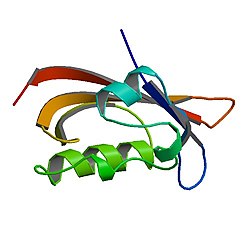
[edit]A detailed atomic structure for hERG based on X-ray crystallography is not yet available, but structures have recently been solved by electron microscopy.[20] In the laboratory the heterologously expressed hERG potassium channel comprises four identical alpha subunits, which form the channel's pore through the plasma membrane. Each hERG subunit consists of 6 transmembrane alpha helices, numbered S1-S6, a pore helix situated between S5 and S6, and cytoplasmically located N- and C-termini. The S4 helix contains a positively charged arginine or lysine amino acid residue at every 3rd position and is thought to act as a voltage-sensitive sensor, which allows the channel to respond to voltage changes by changing conformations between conducting and non-conducting states (called 'gating'). Between the S5 and S6 helices, there is an extracellular loop (known as 'the turret') and 'the pore loop', which begins and ends extracellularly but loops into the plasma membrane; the pore loop for each of the hERG subunits in one channel faces into the ion-conducting pore and is adjacent to the corresponding loops of the three other subunits, and together they form the selectivity filter region of the channel pore. The selectivity sequence, SVGFG, is very similar to that contained in bacterial KcsA channels.[7] Although a full crystal structure for hERG is not yet available, a structure has been found for the cytoplasmic N-terminus, which was shown to contain a PAS domain (aminoacid 26–135) that slows the rate of deactivation.[21]
Genetics
[edit]Loss-of-function mutations in this channel may lead to long QT syndrome (LQT2), while gain-of-function mutations may lead to short QT syndrome. Both clinical disorders stem from ion channel dysfunction (so-called channelopathies) that can lead to the risk of potentially fatal cardiac arrhythmias (e.g., torsades de pointes), due to repolarization disturbances of the cardiac action potential.[18][22] There are far more hERG mutations described for long QT syndrome than for short QT syndrome.[5]
Drug interactions
[edit]This channel is also sensitive to drug binding, as well as decreased extracellular potassium levels, both of which can result in decreased channel function and drug-induced (acquired) long QT syndrome. Among the drugs that can cause QT prolongation, the more common ones include antiarrhythmics (especially Class 1A and Class III), anti-psychotic agents, and certain antibiotics (including quinolones and macrolides).[7]
Although there exist other potential targets for cardiac adverse effects, the vast majority of drugs associated with acquired QT prolongation are known to interact with the hERG potassium channel. One of the main reasons for this phenomenon is the larger inner vestibule of the hERG channel, thus providing more space for many different drug classes to bind and block this potassium channel.[23]
hERG containing channels are blocked by amiodarone, and it does prolong the QT interval, but its multiple other antiarrhythmic effects prevent this from causing torsades de pointes.[24]
Thioridazine causes peculiarly severe QTc prolongation by blocking hERG and was withdrawn by the manufacturer for this reason.
Drug development considerations
[edit]Due to the documented potential of QT-interval-prolonging drugs, the United States Food and Drug Administration issued recommendations for the establishment of a cardiac safety profile during pre-clinical drug development: ICH S7B.[25] The nonclinical evaluation of the potential for delayed ventricular repolarization (QT interval prolongation) by human pharmaceuticals, issued as CHMP/ICH/423/02, adopted by CHMP in May 2005. Preclinical hERG studies should be accomplished in GLP environment.
Naming
[edit]The hERG gene was first named and described in a paper by Jeff Warmke and Barry Ganetzky, then both at the University of Wisconsin–Madison.[16] The hERG gene is the human homolog of the Ether-à-go-go gene found in the Drosophila fly; Ether-à-go-go was named in the 1960s by William D. Kaplan and William E. Trout, III, while at the City of Hope Hospital in Duarte, California. When flies with mutations in the Ether-à-go-go gene are anaesthetised with ether, their legs start to shake, like the dancing at the then popular Whisky a Go Go nightclub in West Hollywood, California.[26]
Interactions
[edit]HERG has been shown to interact with the 14-3-3 epsilon protein, encoded by YWHAE.[27]
Inhibitors
[edit]- Astemizole
- Domperidone
- Sertindole
- Terfenadine
- Vanoxerine (GBR-12909)
See also
[edit]References
[edit]- ^ a b c GRCh38: Ensembl release 89: ENSG00000055118 – Ensembl, May 2017
- ^ a b c GRCm38: Ensembl release 89: ENSMUSG00000038319 – Ensembl, May 2017
- ^ "Human PubMed Reference:". National Center for Biotechnology Information, U.S. National Library of Medicine.
- ^ "Mouse PubMed Reference:". National Center for Biotechnology Information, U.S. National Library of Medicine.
- ^ a b Hedley PL, Jørgensen P, Schlamowitz S, Wangari R, Moolman-Smook J, Brink PA, et al. (November 2009). "The genetic basis of long QT and short QT syndromes: a mutation update". Human Mutation. 30 (11): 1486–1511. doi:10.1002/humu.21106. PMID 19862833. S2CID 19122696.
- ^ Bjerregaard P (August 2018). "Diagnosis and management of short QT syndrome". Heart Rhythm. 15 (8): 1261–1267. doi:10.1016/j.hrthm.2018.02.034. PMID 29501667. S2CID 4519580.
- ^ a b c d Sanguinetti MC, Tristani-Firouzi M (March 2006). "hERG potassium channels and cardiac arrhythmia". Nature. 440 (7083): 463–469. Bibcode:2006Natur.440..463S. doi:10.1038/nature04710. PMID 16554806. S2CID 929837.
- ^ Chiesa N, Rosati B, Arcangeli A, Olivotto M, Wanke E (June 1997). "A novel role for HERG K+ channels: spike-frequency adaptation". The Journal of Physiology. 501 (Pt 2): 313–318. doi:10.1111/j.1469-7793.1997.313bn.x. PMC 1159479. PMID 9192303.
- ^ Overholt JL, Ficker E, Yang T, Shams H, Bright GR, Prabhakar NR (2002). "Chemosensing at the Carotid Body". Oxygen Sensing. Advances in Experimental Medicine and Biology. Vol. 475. pp. 241–248. doi:10.1007/0-306-46825-5_22. ISBN 978-0-306-46367-9. PMID 10849664.
- ^ Arcangeli A (2005). "Expression and Role of hERG Channels in Cancer Cells". The hERG Cardiac Potassium Channel: Structure, Function and Long QT Syndrome. Novartis Foundation Symposia. Vol. 266. pp. 225–32, discussion 232–4. doi:10.1002/047002142X.ch17. ISBN 9780470021408. PMID 16050271.
- ^ Weerapura M, Nattel S, Chartier D, Caballero R, Hébert TE (April 2002). "A comparison of currents carried by HERG, with and without coexpression of MiRP1, and the native rapid delayed rectifier current. Is MiRP1 the missing link?". The Journal of Physiology. 540 (Pt 1): 15–27. doi:10.1113/jphysiol.2001.013296. PMC 2290231. PMID 11927665.
- ^ Abbott GW, Goldstein SA (March 2002). "Disease-associated mutations in KCNE potassium channel subunits (MiRPs) reveal promiscuous disruption of multiple currents and conservation of mechanism". FASEB Journal. 16 (3): 390–400. doi:10.1096/fj.01-0520hyp. PMID 11874988. S2CID 26205057.
- ^ Abbott GW, Sesti F, Splawski I, Buck ME, Lehmann MH, Timothy KW, et al. (April 1999). "MiRP1 forms IKr potassium channels with HERG and is associated with cardiac arrhythmia". Cell. 97 (2): 175–187. doi:10.1016/S0092-8674(00)80728-X. PMID 10219239. S2CID 8507168.
- ^ Jones EM, Roti Roti EC, Wang J, Delfosse SA, Robertson GA (October 2004). "Cardiac IKr channels minimally comprise hERG 1a and 1b subunits". The Journal of Biological Chemistry. 279 (43): 44690–44694. doi:10.1074/jbc.M408344200. PMID 15304481.
- ^ a b Robertson GA, Jones EM, Wang J (2005). "Gating and Assembly of Heteromeric hERG1a/1b Channels Underlying IKr in the Heart". The hERG Cardiac Potassium Channel: Structure, Function and Long QT Syndrome. Novartis Foundation Symposia. Vol. 266. pp. 4–15, discussion 15–8, 44–5. CiteSeerX 10.1.1.512.5443. doi:10.1002/047002142X.ch2. ISBN 9780470021408. PMID 16050259.
- ^ a b Warmke JW, Ganetzky B (April 1994). "A family of potassium channel genes related to eag in Drosophila and mammals". Proceedings of the National Academy of Sciences of the United States of America. 91 (8): 3438–3442. Bibcode:1994PNAS...91.3438W. doi:10.1073/pnas.91.8.3438. PMC 43592. PMID 8159766.
- ^ a b Trudeau MC, Warmke JW, Ganetzky B, Robertson GA (July 1995). "HERG, a human inward rectifier in the voltage-gated potassium channel family". Science. 269 (5220): 92–95. Bibcode:1995Sci...269...92T. doi:10.1126/science.7604285. PMID 7604285.
- ^ a b c Sanguinetti MC, Jiang C, Curran ME, Keating MT (April 1995). "A mechanistic link between an inherited and an acquired cardiac arrhythmia: HERG encodes the IKr potassium channel". Cell. 81 (2): 299–307. doi:10.1016/0092-8674(95)90340-2. PMID 7736582. S2CID 240044.
- ^ Robertson GA (March 2000). "LQT2 : amplitude reduction and loss of selectivity in the tail that wags the HERG channel". Circulation Research. 86 (5): 492–493. doi:10.1161/01.res.86.5.492. PMID 10720408.
- ^ Wang W, MacKinnon R (April 2017). "Cryo-EM Structure of the Open Human Ether-à-go-go-Related K+ Channel hERG". Cell. 169 (3): 422–430.e10. doi:10.1016/j.cell.2017.03.048. PMC 5484391. PMID 28431243.
- ^ Morais Cabral JH, Lee A, Cohen SL, Chait BT, Li M, Mackinnon R (November 1998). "Crystal structure and functional analysis of the HERG potassium channel N terminus: a eukaryotic PAS domain". Cell. 95 (5): 649–655. doi:10.1016/S0092-8674(00)81635-9. PMID 9845367. S2CID 15090987.
- ^ Moss AJ, Zareba W, Kaufman ES, Gartman E, Peterson DR, Benhorin J, et al. (February 2002). "Increased risk of arrhythmic events in long-QT syndrome with mutations in the pore region of the human ether-a-go-go-related gene potassium channel". Circulation. 105 (7): 794–799. doi:10.1161/hc0702.105124. PMID 11854117. S2CID 2616052.
- ^ Milnes JT, Crociani O, Arcangeli A, Hancox JC, Witchel HJ (July 2003). "Blockade of HERG potassium currents by fluvoxamine: incomplete attenuation by S6 mutations at F656 or Y652". British Journal of Pharmacology. 139 (5): 887–898. doi:10.1038/sj.bjp.0705335. PMC 1573929. PMID 12839862.
- ^ "Amiodarone". Drugbank. Retrieved 2019-05-28.
- ^ "S7B Nonclinical Evaluation of the Potential for Delayed Ventricular Repolarization (QT Interval Prolongation) by Human Pharmaceuticals" (PDF). Food and Drug Administration. 6 May 2020.
- ^ Kaplan WD, Trout WE (February 1969). "The behavior of four neurological mutants of Drosophila". Genetics. 61 (2): 399–409. doi:10.1093/genetics/61.2.399. PMC 1212165. PMID 5807804.
- ^ Kagan A, Melman YF, Krumerman A, McDonald TV (April 2002). "14-3-3 amplifies and prolongs adrenergic stimulation of HERG K+ channel activity". The EMBO Journal. 21 (8): 1889–1898. doi:10.1093/emboj/21.8.1889. PMC 125975. PMID 11953308.
Further reading
[edit]- Vincent GM (1998). "The molecular genetics of the long QT syndrome: genes causing fainting and sudden death". Annual Review of Medicine. 49: 263–274. doi:10.1146/annurev.med.49.1.263. PMID 9509262.
- Ackerman MJ (March 1998). "The long QT syndrome: ion channel diseases of the heart". Mayo Clinic Proceedings. 73 (3): 250–269. doi:10.4065/73.3.250. PMID 9511785.
- Taglialatela M, Castaldo P, Pannaccione A, Giorgio G, Annunziato L (June 1998). "Human ether-a-gogo related gene (HERG) K+ channels as pharmacological targets: present and future implications". Biochemical Pharmacology. 55 (11): 1741–1746. doi:10.1016/S0006-2952(98)00002-1. hdl:11566/37313. PMID 9714291.
- Bjerregaard P, Gussak I (February 2005). "Short QT syndrome: mechanisms, diagnosis and treatment". Nature Clinical Practice. Cardiovascular Medicine. 2 (2): 84–87. doi:10.1038/ncpcardio0097. PMID 16265378. S2CID 10125533.
- Gutman GA, Chandy KG, Grissmer S, Lazdunski M, McKinnon D, Pardo LA, et al. (December 2005). "International Union of Pharmacology. LIII. Nomenclature and molecular relationships of voltage-gated potassium channels". Pharmacological Reviews. 57 (4): 473–508. doi:10.1124/pr.57.4.10. PMID 16382104. S2CID 219195192.


