CXCL1
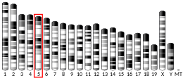
The chemokine (C-X-C motif) ligand 1 (CXCL1) is a small peptide belonging to the CXC chemokine family that acts as a chemoattractant for several immune cells, especially neutrophils[5][6] or other non-hematopoietic cells to the site of injury or infection and plays an important role in regulation of immune and inflammatory responses. It was previously called GRO1 oncogene, GROα, neutrophil-activating protein 3 (NAP-3) and melanoma growth stimulating activity, alpha (MGSA-α). CXCL1 was first cloned from a cDNA library of genes induced by platelet-derived growth factor (PDGF) stimulation of BALB/c-3T3 murine embryonic fibroblasts and named "KC" for its location in the nitrocellulose colony hybridization assay.[7] This designation is sometimes erroneously believed to be an acronym and defined as "keratinocytes-derived chemokine". Rat CXCL1 was first reported when NRK-52E (normal rat kidney-52E) cells were stimulated with interleukin-1β (IL-1β) and lipopolysaccharide (LPS) to generate a cytokine that was chemotactic for rat neutrophils, cytokine-induced neutrophil chemoattractant (CINC).[8] In humans, this protein is encoded by the gene CXCL1 [9] and is located on human chromosome 4 among genes for other CXC chemokines.[10]
Structure and expression
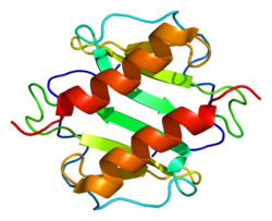
[edit]CXCL1 exists as both monomer and dimer and both forms are able to bind chemokine receptor CXCR2.[11] However, CXCL1 chemokine is able to dimerize only at higher (micromolar) concentrations and its concentrations are only nanomolar or picomolar upon normal conditions, which means that the form of WT CXCL1 is more likely monomeric while dimeric CXCL1 is present only during infection or injury. CXCL1 monomer consists of three antiparallel β-strands followed by C- terminal α-helix and this α-helix together with the first β-strand are involved in forming a dimeric globular structure.[12]
Upon normal conditions, CXCL1 is not expressed constitutively. It's produced by a variety of immune cells such as macrophages, neutrophils and epithelial cells,[13][14] or Th17 population. Moreover, its expression can be also induced indirectly by IL-1, TNF-α or IL-17 produced again by Th17 cells [15] and is triggered mainly by activation of NF-κB or C/EBPβ signaling pathways predominantly involved in inflammation and leading to production of other inflammatory cytokines.[15]
Function
[edit]CXCL1 has a potentially similar role as interleukin-8 (IL-8/CXCL8). After binding to its receptor CXCR2, CXCL1 activates phosphatidylinositol-4,5-bisphosphate 3-kinase-γ (PI3Kγ)/Akt, MAP kinases such as ERK1/ERK2 or phospholipase-β (PLCβ) signaling pathways. CXCL1 is expressed at higher levels during inflammatory responses thus contributing to the process of inflammation.[16] CXCL1 is also involved in the processes of wound healing and tumorigenesis.[17][18][19]
Role in cancer
[edit]CXCL1 has a role in angiogenesis and arteriogenesis [20] and thus has been shown to act in the process of tumor progression. The role of CXCL1 was described by several studies in the development of various tumors, such as breast cancer, gastric and colorectal carcinoma or lung cancer.[21][22][23] Also, CXCL1 is secreted by human melanoma cells, has mitogenic properties and is implicated in melanoma pathogenesis.[24][25][26]
Role in nervous system and sensitization
[edit]CXCL1 plays a role in spinal cord development by inhibiting the migration of oligodendrocyte precursors.[11] CXCR2 receptor for CXCL1 is expressed in the brain and spinal cord by neurons and oligodendrocytes and during CNS pathologies such as Alzheimer's disease, multiple sclerosis and brain injury also by microglia. An initial study in mice showed evidence that CXCL1 decreased the severity of multiple sclerosis and may offer a neuro-protective function.[27] On the other hand, on the periphery, CXCL1 contributes to the release of prostaglandins and thus causes increased sensitivity to pain and drives nociceptive sensitization via recruitment of neutrophils to the tissue. Phosphorylation of ERK1/ERK2 kinases and activation of NMDA receptors leads to transcription of genes inducing chronic pain, such as c-Fos or cyclooxygenase-2 (COX-2).[16]
References
[edit]- ^ a b c GRCh38: Ensembl release 89: ENSG00000163739 – Ensembl, May 2017
- ^ a b c GRCm38: Ensembl release 89: ENSMUSG00000058427 – Ensembl, May 2017
- ^ "Human PubMed Reference:". National Center for Biotechnology Information, U.S. National Library of Medicine.
- ^ "Mouse PubMed Reference:". National Center for Biotechnology Information, U.S. National Library of Medicine.
- ^ Moser B, Clark-Lewis I, Zwahlen R, Baggiolini M (May 1990). "Neutrophil-activating properties of the melanoma growth-stimulatory activity". The Journal of Experimental Medicine. 171 (5): 1797–1802. doi:10.1084/jem.171.5.1797. PMC 2187876. PMID 2185333.
- ^ Schumacher C, Clark-Lewis I, Baggiolini M, Moser B (November 1992). "High- and low-affinity binding of GRO alpha and neutrophil-activating peptide 2 to interleukin 8 receptors on human neutrophils". Proceedings of the National Academy of Sciences of the United States of America. 89 (21): 10542–10546. Bibcode:1992PNAS...8910542S. doi:10.1073/pnas.89.21.10542. PMC 50375. PMID 1438244.
- ^ Cochran BH, Reffel AC, Stiles CD (July 1983). "Molecular cloning of gene sequences regulated by platelet-derived growth factor". Cell. 33 (3): 939–947. doi:10.1016/0092-8674(83)90037-5. PMID 6872001. S2CID 38719612.
- ^ Watanabe K, Kinoshita S, Nakagawa H (June 1989). "Purification and characterization of cytokine-induced neutrophil chemoattractant produced by epithelioid cell line of normal rat kidney (NRK-52E cell)". Biochemical and Biophysical Research Communications. 161 (3): 1093–1099. doi:10.1016/0006-291X(89)91355-7. PMID 2662972.
- ^ Haskill S, Peace A, Morris J, Sporn SA, Anisowicz A, Lee SW, et al. (October 1990). "Identification of three related human GRO genes encoding cytokine functions". Proceedings of the National Academy of Sciences of the United States of America. 87 (19): 7732–7736. Bibcode:1990PNAS...87.7732H. doi:10.1073/pnas.87.19.7732. PMC 54822. PMID 2217207.
- ^ Richmond A, Balentien E, Thomas HG, Flaggs G, Barton DE, Spiess J, et al. (July 1988). "Molecular characterization and chromosomal mapping of melanoma growth stimulatory activity, a growth factor structurally related to beta-thromboglobulin". The EMBO Journal. 7 (7): 2025–2033. doi:10.1002/j.1460-2075.1988.tb03042.x. PMC 454478. PMID 2970963.
- ^ a b Tsai HH, Frost E, To V, Robinson S, Ffrench-Constant C, Geertman R, et al. (August 2002). "The chemokine receptor CXCR2 controls positioning of oligodendrocyte precursors in developing spinal cord by arresting their migration". Cell. 110 (3): 373–383. doi:10.1016/S0092-8674(02)00838-3. PMID 12176324. S2CID 16880392.
- ^ Ravindran A, Sawant KV, Sarmiento J, Navarro J, Rajarathnam K (April 2013). "Chemokine CXCL1 dimer is a potent agonist for the CXCR2 receptor". The Journal of Biological Chemistry. 288 (17): 12244–12252. doi:10.1074/jbc.m112.443762. PMC 3636908. PMID 23479735.
- ^ Iida N, Grotendorst GR (October 1990). "Cloning and sequencing of a new gro transcript from activated human monocytes: expression in leukocytes and wound tissue". Molecular and Cellular Biology. 10 (10): 5596–5599. doi:10.1128/mcb.10.10.5596. PMC 361282. PMID 2078213.
- ^ Becker S, Quay J, Koren HS, Haskill JS (March 1994). "Constitutive and stimulated MCP-1, GRO alpha, beta, and gamma expression in human airway epithelium and bronchoalveolar macrophages". The American Journal of Physiology. 266 (3 Pt 1): L278–L286. doi:10.1152/ajplung.1994.266.3.L278. PMID 8166297.
- ^ a b Ma K, Yang L, Shen R, Kong B, Chen W, Liang J, et al. (March 2018). "Th17 cells regulate the production of CXCL1 in breast cancer". International Immunopharmacology. 56: 320–329. doi:10.1016/j.intimp.2018.01.026. PMID 29438938. S2CID 3568978.
- ^ a b Silva RL, Lopes AH, Guimarães RM, Cunha TM (September 2017). "CXCL1/CXCR2 signaling in pathological pain: Role in peripheral and central sensitization". Neurobiology of Disease. 105: 109–116. doi:10.1016/j.nbd.2017.06.001. PMID 28587921. S2CID 4916646.
- ^ Devalaraja RM, Nanney LB, Du J, Qian Q, Yu Y, Devalaraja MN, Richmond A (August 2000). "Delayed wound healing in CXCR2 knockout mice". The Journal of Investigative Dermatology. 115 (2): 234–244. doi:10.1046/j.1523-1747.2000.00034.x. PMC 2664868. PMID 10951241.
- ^ Haghnegahdar H, Du J, Wang D, Strieter RM, Burdick MD, Nanney LB, et al. (January 2000). "The tumorigenic and angiogenic effects of MGSA/GRO proteins in melanoma". Journal of Leukocyte Biology. 67 (1): 53–62. doi:10.1002/jlb.67.1.53. PMC 2669312. PMID 10647998.[permanent dead link]
- ^ Owen JD, Strieter R, Burdick M, Haghnegahdar H, Nanney L, Shattuck-Brandt R, Richmond A (September 1997). "Enhanced tumor-forming capacity for immortalized melanocytes expressing melanoma growth stimulatory activity/growth-regulated cytokine beta and gamma proteins". International Journal of Cancer. 73 (1): 94–103. doi:10.1002/(SICI)1097-0215(19970926)73:1<94::AID-IJC15>3.0.CO;2-5. PMID 9334815.
- ^ Vries MH, Wagenaar A, Verbruggen SE, Molin DG, Dijkgraaf I, Hackeng TH, Post MJ (April 2015). "CXCL1 promotes arteriogenesis through enhanced monocyte recruitment into the peri-collateral space". Angiogenesis. 18 (2): 163–171. doi:10.1007/s10456-014-9454-1. PMID 25490937. S2CID 52835567.
- ^ Chen X, Jin R, Chen R, Huang Z (2018-02-01). "Complementary action of CXCL1 and CXCL8 in pathogenesis of gastric carcinoma". International Journal of Clinical and Experimental Pathology. 11 (2): 1036–1045. PMC 6958037. PMID 31938199.
- ^ Hsu YL, Chen YJ, Chang WA, Jian SF, Fan HL, Wang JY, Kuo PL (August 2018). "Interaction between Tumor-Associated Dendritic Cells and Colon Cancer Cells Contributes to Tumor Progression via CXCL1". International Journal of Molecular Sciences. 19 (8): 2427. doi:10.3390/ijms19082427. PMC 6121631. PMID 30115896.
- ^ Spaks A (April 2017). "Role of CXC group chemokines in lung cancer development and progression". Journal of Thoracic Disease. 9 (Suppl 3): S164–S171. doi:10.21037/jtd.2017.03.61. PMC 5392545. PMID 28446981.
- ^ Anisowicz A, Bardwell L, Sager R (October 1987). "Constitutive overexpression of a growth-regulated gene in transformed Chinese hamster and human cells". Proceedings of the National Academy of Sciences of the United States of America. 84 (20): 7188–7192. Bibcode:1987PNAS...84.7188A. doi:10.1073/pnas.84.20.7188. PMC 299255. PMID 2890161.
- ^ Richmond A, Thomas HG (February 1988). "Melanoma growth stimulatory activity: isolation from human melanoma tumors and characterization of tissue distribution". Journal of Cellular Biochemistry. 36 (2): 185–198. doi:10.1002/jcb.240360209. PMID 3356754. S2CID 10674236.
- ^ Dhawan P, Richmond A (July 2002). "Role of CXCL1 in tumorigenesis of melanoma". Journal of Leukocyte Biology. 72 (1): 9–18. doi:10.1189/jlb.72.1.9. PMC 2668262. PMID 12101257.
- ^ Omari KM, Lutz SE, Santambrogio L, Lira SA, Raine CS (January 2009). "Neuroprotection and remyelination after autoimmune demyelination in mice that inducibly overexpress CXCL1". The American Journal of Pathology. 174 (1): 164–176. doi:10.2353/ajpath.2009.080350. PMC 2631329. PMID 19095949.
External links
[edit]- Human CXCL1 genome location and CXCL1 gene details page in the UCSC Genome Browser.


