Wikipedia:Wiki Science Competition 2023 in the United States/Results
The 2023 Wiki Science Competition in the United States announces its national finalists, all of which will advance to the international phase of the competition. The Wiki Science Competition is an international science photography competition first organized in Estonia, expanding in 2015 to all of Europe, and in 2017 to the rest of the world. The U.S. branch of the competition has been organized by Wikimedia District of Columbia since 2017.
Participants uploaded their submissions during November and December 2023. The U.S. competition received 460 contributions across seven categories. These included 246 images of wildlife, 39 microscopy images, 25 astronomy images, 12 images of people in science, 4 non-photographic and video submissions, 98 general category images, and 36 images as part of sets. The jury selected up to seven winners in each category to represent the United States at the international level. Of the 32 finalists, only 5 had previous uploads to Wikimedia Commons. In addition, the panels selected eight submissions as "Jury's Choice" awards, each of which receives a $250 prize from Wikimedia District of Columbia.
Thanks to our volunteer judges for their efforts. The jury for the U.S. competition is listed below.
Jury's Choice Award winners
[edit] |
Association of Widefield Astrophotographers, consisting of Braxton Townsend, Ryder Cobean, Jacob Newbould, Jayton Nelson, Uri Darom, and Mikołaj Wadowski, is honored for Andromeda and integrated flux nebulae, showing the famous Andromeda Galaxy and its extensive outer halo, revealing various faint structures, some very recently discovered. The data is mostly from high schoolers and college students, proving that very expensive equipment and dark skies aren't required to create unique images of faint objects.
We all started astrophotography for unique reasons. However, what probably brought us all together was our desire to explore the universe far beyond our own planet. Astrophotography is the only hobby that allows one to reach millions of light years from home, and look back millions of years in the past. Astronomy offers a perspective on the world that is truly unique to itself. We created the Association for Widefield Astrophotographers to serve one goal: to create unique, never before seen perspectives on popular objects in the sky, and to do so with affordable gear that is available to even the up and coming photographer. We realized that any disadvantage, including the limitations imposed by cheaper gear or light polluted skies, can be overcome through collaboration. To do this, we assembled an international group of young astronomers, all using the same affordable gear. We initially set our sights on the Andromeda Galaxy as our first target due to its popularity. As the largest, brightest galaxy in the sky, outside of our own Milky Way, the Andromeda Galaxy (also known as Messier 31 or M31) is one of the most photographed deep sky objects ever. However, we knew from some recent discoveries that there was a lot more hiding in this seemingly familiar area of the sky. And so we got to work; six people from around the world spent night after night taking images of this galaxy. Overall, we collected 100 hours of data. The next challenge was combining everyone’s photographs together. From varying skies, to locations, to gear, merging everyone’s photos proved to be a challenge in and of itself. We think the work paid off though; after processing the image, we were able to reveal everything we had hoped for, from the faint red Hydrogen Alpha clouds surrounding the galaxy to the newly discovered Oxygen III nebula seemingly resting above it. We hope this photograph can serve as a reminder to challenge what we think is normal; there is always more waiting to be discovered, both in astrophotography and beyond. Moreso, we hope that this project can inspire people to enter astrophotography without the impression that expensive equipment and dark skies are needed to get great results. Patience, dedication, and collaboration are all that is required. |
 |
Joan Dudney is honored for Whitebark pine forest, showing a forest dominated by whitebark pine in the eastern Sierra Nevada. Whitebark pine is imperiled by climate change, bark beetles and disease, and recently listed as endangered due to its rapid decline throughout North America. It provides critical habitat for a diversity of wildlife, including grizzly bears, birds, and squirrels, and regions with high mortality experience dramatic shifts in water and nutrient cycling.
I am an assistant professor at UC Santa Barbara and my research focuses on understanding global change impacts on forest ecosystems. One of my projects seeks to understand how complex, interacting drivers of tree mortality are affecting the health of whitebark pine in the Sierra Nevada. Whitebark pine is a particularly unique and important tree species that provides critical ecosystem services and habitat for iconic wildlife. Unfortunately, due to a combination of threats, including an invasive tree disease, mountain pine beetle, and drought, we are seeing unprecedented mortality throughout its range, which led to its recent listing as "Threatened" under the Endangered Species Act. This photo captures a region of the whitebark pine-dominated forests in the Sierra Nevada that is still relatively intact. It also happens to be an incredibly scenic place that exemplifies why the Sierra Nevada is so deeply loved by recreationists and photographers! I captured this photo on one of my research trips to this site. I was conducting a field study that extracted stable isotopes and tree rings to understand the long-term effects of extreme drought on whitebark pine growth and photosynthesis. The swift-flowing, clear water surrounded by whitebark pine forests and perfectly chiseled mountains remind me of why I do this work and how important it is to protect the remaining healthy stands—the foundation of these awe-inspiring ecosystems. My research in the Sierra has also inspired me to learn more about photography, so I can share the beauty and rapid change in these forests with the public. I started my photography using an iphone when I was a PhD student, and this has slowly turned into a hobby that keeps me hopeful in the face of the forest mortality that I witness every year. The rapid decline of Sierran forests is a constant reminder that we have to make pretty dramatic changes as a society to mitigate our negative impacts and steward these lands effectively. |
 |
Mirai Kambayashi is honored for Crown shyness, a feature observed in some tree species, in which the crowns of fully stocked trees do not touch each other, instead forming a canopy with channel-like gaps.
I am a high-school student who loves to observe and research natural phenomena. My way of sharing this passion with others is through digital media. This way, I can let others see what I see directly through my lens. I try to invite discussion and hypotheses through my media. This competition was a perfect way to do so, since crown shyness is a niche phenomenon with lots of potential; it invites ideas about physical communication between the same species of trees. As I was walking back from the store, I saw this phenomenon. I took this picture at Koganei Park in Tokyo, Japan. |
 |
Saidelsky is honored for Fritz Goro at work. Fritz Goro was a premier science photographer in the United States and the inventor of macrophotography. This is a 1972 photograph in the laboratory of Drs. Hubel and Weisel at Harvard Medical School, where he was hired to illustrate their investigations of vision in monkeys, for which they won the Nobel Prize in 1981.
In 1972, I had graduated from MIT and was working in a neurobiology lab. I was hired by him to carry his equipment on assignment to the laboratory of Drs. Hubel and Weisel. Since I was deciding between a career as a neurobiologist or photographer, working with him provided me with the opportunity to decide. |
  |
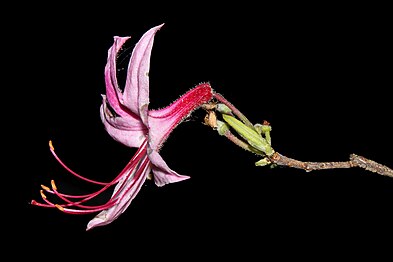
Lily Rice is honored for an image set of Imperiled and vulnerable flora of Ohio. Some of these plants are limited to a single habitat in the state. For example, Platanthera ciliaris is limited to two populations on one of the rarest habitat types in the world, oak savannas, while the the Ohio population of the diminutive orchid Cypripedium candidum is limited to a single remnant prairie near Sandusky.
In the wake of the anthropocene, plant blindness is a malady that impacts us all. My goal as a scientific educator is to not only detail the beauty of complex, botanical life, but also assist others in understanding the critical role that plants serve as the basis of all life on land. Plant adaptations to their environments are more subtle, but equally (if not moreso) fascinating to animal traits. My own interest in botany began with an accidental poisoning that unlocked a rapid appreciation for the little wonders that plants are. Quickly, I wanted to absorb as much inofrmation as I could on native plants and over time, I began to share this information to anyone who would listen. I formed an Instagram page and website—both of which are titled, "Watching Plant Sex in the Woods," after the act botanists spend their time doing—where I primarily educate lay-people on whatever native plant subject I desire. Quickly, I decided my phone camera was inadequate to capture the detail I wanted so I decided to pick up the expensive hobby of photography. Flash forward to my image set presented here and you can see two different styles and two different cameras. My 'black box' series was a seasonal study on highlighting floral morphology by dropping the background out of the image, which gives a focused appreciation on floral symmetry, purpose, and function. These images were shot on a Canon Rebel T7i body and a special black box made from an arborist's throw cube. In addition, the image titled, "Fen Familiars," was captured on the same camera in one of my favorite habitats in the world, a fen. Finally, my more recent images were shot on a Nikon Z7 body—those photos are of three uncommon species in Ohio, all of which were on my must see list. The most important species for me was my botanical white whale, Platanthera ciliaris, which is found only in two spots in Ohio. The ornate, orange frills are beautiful to behold yes, but serve a likely reproductive purpose as footholds for their moth pollinators! As a photographer, I must admit that I am rather lazy. My primary goal is to see species in habitat and learn as much as I can. Most times I forget to even take photos as I am too enthralled with the subject. All of this being said, I submit my photos to competitions, such as the Wiki Science Competition, to help others realize the importance of plants and the subtle, but incredible, adaptations they wield. |
 |
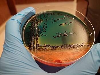
Amanda Toney is honored for Stool culture on agar. The stool culture grown on Hektoen enteric agar exhibits two differential traits for two different microbes: the orange color indicates fermentation of lactose, likely from E. coli, while the black color is from the microbial production of hydrogen sulfide, and is often seen in Salmonella spp.
I took this photo on April 13, 2022 while I was taking my first pathogenic microbiology class in the medical laboratory technology program at Rose State College. We didn't always have time to fill out our lab reports and worksheets in the lab, and so my classmates and I made a habit of taking pictures of our cultures with our phones and doing our homework later based on them. I took a lot of pictures of this particular culture because, even though it was grown of liquid feces and composed almost exclusively of pathogens, I couldn't help but think it was pretty and reminded me of a sunset. I think there's beauty to be found in even the more "disgusting" aspects of biology. I think that the infinite complexity of everything is worth marveling at, despite the smell. I love to take pictures, especially in the lab and in nature. My grandfather was a photographer and an inventor, and some part of me feels like I ought to continue that legacy somehow. I also find great joy in sharing the things I learn in my studies and work with others, making science a little more accessible and interesting to the people who otherwise don't think about it. I'm studying for my bachelor's in biomedical science, now. It's been slow because I have to balance school with working full-time as an medical laboratory technician in what I can only describe as a STAT lab, mostly limited to routine chemistry, hematology, and urinalysis testing from low-acuity patients (usually). I still have a fondness for and interest in microbiology, but I've heard nothing but warnings from other lab techs against working for the three major companies in the Oklahoma City metro that still do substantial microbiology testing in their laboratories; those who've worked for them get a scared look in their eye when I ask how it is to work there, usually followed by a firm, "Don't." TL;DR: bacteria are cool and corporate greed gutted the microbiology field. Sad! |
 |
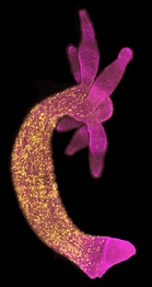
Ben D. Cox is honored for Stem cell distribution in Hydra vulgaris. A population of interstitial stem cells (yellow) gives rise to neurons, gland cells, gametes, and stinging cells. These stem cells are located only in the the outer tissue layer of the body column, but must make it to locations in the entire animal through the extracellular matrix (magenta).
I became interested in how animals grow when studying zebrafish eye development as an undergraduate at the University of Texas. The field of developmental biology relies on seeing changes in organisms at the cellular and tissue levels, and I developed a knack for using a confocal microscope to take fluorescent images. Perhaps because I am naturally more of a verbal/written learner, I was attracted to the challenge of thinking about biological questions through visual data. My research deals with how Hydra is able to regenerate after serious injury, and specifically how these stem cells get where they need to go—including moving through a layer of collagen—to replenish lost cell populations. However, I first wanted to understand how Hydra’s stem cells are distributed throughout its body in an uninjured state, hence the perfectly intact animal depicted in this image. I decided to submit this image to the Wiki Science Competition because much of scientific research, especially biomedical research, focuses on a small number of model organisms like mice and flies. Because I study an animal that is not among those models, I want to reach out to the public and show them how much exciting biology can be done all throughout the tree of life. The Wiki Science Competition is a great avenue for people working inside or outside the scientific mainstream, as professionals or amateurs, to communicate the beauty of their research. |
 |
David S. Goodsell is honored for Bacteriophage T4 infection lifecycle. At left, a bacteriophage is injecting its DNA genome into an E. coli cell. At center, the bacteriophage has taken over the cell, destroying the cellular DNA and forcing the cell to make many new copies of itself. At right, the bacteriophage causes the cell to burst, releasing several hundred new bacteriophages.
My illustrations depict a level of biological scale that is largely invisible to experiment, making artistic visualization a central tool for intuition and understanding. My illustrations integrate information from many experimental sources, building up a picture of what we might see if we could look into a living cell and view it at the molecular level. To see additional examples of my watercolor and digital illustrations, visit https://pdb101.rcsb.org/sci-art/goodsell-gallery |
All finalists
[edit]All 45 U.S. finalists are displayed below. The full results gallery contains more information about the winning photographs, including the complete winning image sets.
-
Andromeda and integrated flux nebulae by Association of Widefield Astrophotographers
-
Crown shyness by Mirai Kambayashi
-
Fritz Goro at work by Saidelsky
-
Imperiled and vulnerable flora of Ohio (set) by Lily Rice
-
Stool culture on agar by Amanda Toney
-
Stem cell distribution in Hydra vulgaris by Ben D. Cox
-
Bacteriophage T4 infection lifecycle by David S. Goodsell
-
2023 annular eclipse by Dpickd1
-
Whitebark pine forest by Treelove776
-
Vortex draining by Wlwiener
-
Amino acid birefringence (set) by Aw1792300
-
Ice core drill head by Kendrick15435
-
Gorgonilla spherules by Hermannbermudez
-
Turbulence stretching orientation map by Terry Brannigan
-
Gamma cas nebula by Ram Samudrala
-
Brown marmorated stink bug by Hyllir
-
Antarctic snow pit by Kendrick15435
-
Nevis linear particle accelerator (set) by CUBIST DEBRIS
-
Fagradalsfjall volcanic eruption by Yuo7si
-
Human brain cell organoid by Nreis1
-
Solutions to characteristic polynomials of degree 7 by Trra02
-
Ram Samudrala
-
Gator reflections by Sam D. Hamilton
-
Scientist examines an ice core by Kendrick15435
-
Crystals in Song Dynasty glaze (set) by Chandra L. Reedy
-
Time travelling 500 million years back by Shibajyotidas
-
Flounder larva by Taeylenol
-
Starlink satellite over Mt. Rainier by WhatWeGetFromThisAdventure
-
Bamboo grove by Yuo7si
-
Digging through a million years of history by Shibajyotidas
-
Vitamin C birefringence (set) by Aw1792300
-
Cloud-to-ground lightning strike by Phiteros
-
Crystals in Song Dynasty glaze by Chandra L. Reedy
-
Lion Nebula of Cepheus by Ram Samudrala
-
Female lion showing teeth by Thecodemachine
-
WAIS Divide ice core by Kendrick15435
-
Fallout from Chernobyl (set) by Yuo7si
-
Nature wins by LittleLeafSheep
-
Posterior end of a nematode by Nemataslg
-
Perseid meteor and Andromeda by Phiteros
-
Pacific sea nettle by Ashley98lee
-
Randall Munroe at Strange Loop by Σ
-
Wildlife of Uganda (set) by Thecodemachine
-
Wind turbines in fog by Phiteros
-
Deer sperm under fluorescence microscopy by Theprevetvikng
Jury
[edit]Thanks to our volunteer judges for their efforts. The jury for the U.S. competition is:
- John P. Sadowski, Wikimedia District of Columbia (coordinator)
- Annie Rauwerda, Depths of Wikipedia creator
- Ben Inouye, 2021 U.S. Jury's Choice Prize and International Runner-Up for "Milky Way at Joshua Tree"
- Callan Carpenter, 2019 U.S. Jury's Choice Prize and International Runner-Up for "Killer whales hunting a seal"
- Jake Saunders, 2019 U.S. Jury's Choice Prize for "Earthworm head"
- Jamie Flood, Wikipedian-in-Residence at National Agricultural Library
- Jeremy Axelrod, 2019 U.S. Jury's Choice Prize and International QSORT Prize for "See the light"
- Kevin Payravi, Wikimedia District of Columbia
- Laura Soito, Associate Dean for Content and Discovery, University of Massachusetts Amherst
- Michael Adler, 2017 and 2019 U.S. Jury's Choice Prize and International Runner-Up for "Total Solar Eclipse" and "Jupiter's South Polar Region"
- Natalie Carrigan, 2019 International Runner-Up for "3D projection of a Patiria miniata bipinnaria"
- Tom Wagner, 2017 International Runner-Up for "Birefringent Water Ice"
- Verne Lehmberg, 2021 U.S. Jury's Choice Prize and International Runner-Up for "Sticky geranium anther"


