Wikipedia:Featured picture candidates/Wrist and hand deeper palmar dissection-en.svg
Appearance

- Reason
- High quality and great value to the encyclopedia
- Articles this image appears in
- Hand
- Creator
- Wilfredor

- Support as nominator --Libertad0 ॐ (talk) 19:48, 29 March 2009 (UTC)
- Support assuming nobody finds anything glaringly wrong about the diagram. Certainly of good quality and fairly easy to understand. Caption is a big unwieldly though. Diliff | (Talk) (Contribs) 08:03, 30 March 2009 (UTC)
- Comment. Legend in the image, or the caption, should indicate whether the palm is up or down-- it's not immediately obvious. Spikebrennan (talk) 13:22, 30 March 2009 (UTC)
- Strong Support - pending resolution of spike's concerns and confirmation of accuracy, this is an incredible image - a brilliant use of SVG and one of the finest diagrams on Wikipedia. —Vanderdecken∴ ∫ξφ 16:44, 30 March 2009 (UTC)
- Support nice diagram, very educational—Chris! ct 22:40, 30 March 2009 (UTC)
- Strong Support, if accuracy is confirmed by participants from WP:Medicine (who have been informed). An excellent image. Mostlyharmless (talk) 03:38, 31 March 2009 (UTC)
- Thank you for your message at WP:Med. I have added a comment below. Snowman (talk) 10:51, 31 March 2009 (UTC)
- Comments this is an outstanding image, but I have some concerns:
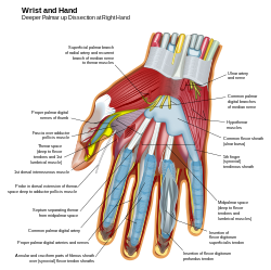
- Why is the fascia that overlies the adductor pollicis so opaque? It obscures the fact that the underlying structure, a muscle, is like the other muscles, and that its fibers are almost perpendicular to the others (hence its functional importance for our opposable thumb).
- Linked to my previous comment - the path of the probe, which is supposedly deep to the adductor pollicis muscle, is unclear - is that probe more than a distraction?
- Shouldn't "anular" be "annular"?
- Is this an opportunity to indicate the stuctures passing through the carpal tunnel?
- My Strong support for this beautiful and technically outstanding image is mitigated by these concerns. --Scray (talk) 04:54, 31 March 2009 (UTC)
- Oppose. Overly medical diagram of limited use to most Wikipedia users. This is basically showing a dissection, complete with a probe, when we would be better served by a more common usage illustration of the hand (who below university level medical students would be using 90% of this terminology?). The general description in the caption doesn't gel with the medical terminology included on the diagram itself. Also completely lacking in sources, which I thought we had agreed was required for diagrams due to concerns about WP:OR. While the diagram itself seems well enough done, I don't think it's serving the target audience well. --jjron (talk) 06:47, 31 March 2009 (UTC)
Oppose: It think the author is potentially a talented illustrator, but I think that the hand image is little use to an anatomist and might confuse a general reader. I do not know why the radial artery is not labelled. I think that too many of the connecting nerves and arteries have been cut out; see File:Gray817.png. I do not know why the anastomosis between the radial artery and the ulnar artery is not shown, which is one of the most important things that needs to be shown here; see File:Gray815.png. The insertion of flexor digitonum superficialis tendon does not show its mechanical arrangement, which is another one of the key features that needs to be shown. I think the common flexor sheath extends further down the hand than illustrated; see File:Gray423.png. Just to show it is there, I might have put in the radial nerve as it innervates the web between the thumb and index finger and also parts of the back of the hand and wrist (with some variability). I think that is would be better to do separate diagrams of the arteries, nerves, muscles with tendons and bones. The diagram should say that it is the anterior aspect (palm up) of a right hand, and I think that all of the structures passing through the carpel tunnel need labelling.I think that the omissions make this image potentially misleading, and, in my opinion, should definitely not be a featured image.I think the caption needs rewriting too. Snowman (talk) 10:32, 31 March 2009 (UTC)
- I don't agree with the strongly negative tone of this comment, for the following reasons: it's a judgment call regarding the balance between satisfying an audience that sits somewhere between but far from the extremes of anatomist versus completely uneducated reader (I'll admit that I'm closer to the former than the latter); this is a deep dissection, and one simply has to make choices about the structures retained (e.g. loss of the palmar arterial arcades); Gray's 1901 edition is not a gold standard against which to measure other illustrations - it's useful and available, but has its limits; I agree with the suggestions regarding labeling and the caption. Separate illustrations of types of structures would be useful, but this sort of combined image helps integrate concepts, too. --Scray (talk) 12:16, 31 March 2009 (UTC)
- I think the omission of the anastomosis in itself would fail this in becoming a featured image with the current caption. It might be better, if all the smaller nerves and arteries were removed and it became an illustration of the muscles, tendons, and tendon sheaths. It would be more understandable, if the caption was helpful and said that important sections of arteries and nerves have been removed. Please list any faults that you have found on the Gray's illustrations that I have linked. Snowman (talk) 13:56, 31 March 2009 (UTC)
- Several comments have been on about the caption, so I have rewritten it and I am sure that it could be improved further. I would pass the image as a featured image with a suitable caption which says that arteries and nerves have been "cut away", which I think the new modified caption does. Snowman (talk) 15:15, 31 March 2009 (UTC)
- I also think that it would be worth a dedicated label for one or more of the lumbrical muscles. Snowman (talk) 22:47, 31 March 2009 (UTC)
- I don't agree with the strongly negative tone of this comment, for the following reasons: it's a judgment call regarding the balance between satisfying an audience that sits somewhere between but far from the extremes of anatomist versus completely uneducated reader (I'll admit that I'm closer to the former than the latter); this is a deep dissection, and one simply has to make choices about the structures retained (e.g. loss of the palmar arterial arcades); Gray's 1901 edition is not a gold standard against which to measure other illustrations - it's useful and available, but has its limits; I agree with the suggestions regarding labeling and the caption. Separate illustrations of types of structures would be useful, but this sort of combined image helps integrate concepts, too. --Scray (talk) 12:16, 31 March 2009 (UTC)
- Support. Firstly, I am amazed at the detail and skill of this SVG, really well done. Secondly I notice that the numbered version is featured on commons (added on right). This potentially could be a more useful image for article space where the text on the text version is likely to be too small unless viewed in full, this would also allow a better caption and variable labelling perhaps with links and medical and lay terms where appropriate.
- This would be a strong support if some clear indication is made of it being palm up; I would prefer the radial artery to also be labelled and perhaps the anastomosis to be present but appreciate this is a great image without this. |→ Spaully₪† 14:14, 31 March 2009 (GMT)
- I have notified the author on commons and perhaps he can attend to any labelling minor fixes. Snowman (talk) 15:25, 31 March 2009 (UTC)
- Good day. I have been reading all your comments, and I am ready to correct these errors. I have problems to identify radial artery. I am not a doctor and my source, Dr. Who has decided to help is not anonymous. Thank's --Libertad0 ॐ (talk) 18:21, 31 March 2009 (UTC)
- Hi, 18 points to the ulnar artery. The radial artery is the red one on the other side of the wrist about the same diameter. There is only a small bit visible before it is cut off. Next to the radial artery are two tendons and in the middle of the wrist there is the median nerve, the large yellow one, which could be labelled median nerve. There are two more tendons to the right and another red tube the ulnar artery, also worth labelling. Snowman (talk) 20:19, 31 March 2009 (UTC)
- A minor point (and it may be variable) but it is the ulnar artery - with an 'r'. It would be good to label the median nerve as you say. |→ Spaully₪† 21:22, 31 March 2009 (GMT)
- Whoops, "ulnar" is the correct spelling. Ulna is a bone. I have corrected the typing above to reduce confusion. Snowman (talk) 22:09, 31 March 2009 (UTC)
- A minor point (and it may be variable) but it is the ulnar artery - with an 'r'. It would be good to label the median nerve as you say. |→ Spaully₪† 21:22, 31 March 2009 (GMT)
- Hi, 18 points to the ulnar artery. The radial artery is the red one on the other side of the wrist about the same diameter. There is only a small bit visible before it is cut off. Next to the radial artery are two tendons and in the middle of the wrist there is the median nerve, the large yellow one, which could be labelled median nerve. There are two more tendons to the right and another red tube the ulnar artery, also worth labelling. Snowman (talk) 20:19, 31 March 2009 (UTC)
- Good day. I have been reading all your comments, and I am ready to correct these errors. I have problems to identify radial artery. I am not a doctor and my source, Dr. Who has decided to help is not anonymous. Thank's --Libertad0 ॐ (talk) 18:21, 31 March 2009 (UTC)
- I have notified the author on commons and perhaps he can attend to any labelling minor fixes. Snowman (talk) 15:25, 31 March 2009 (UTC)
- Support. Excellent vector image. Even if I don't feel like rehearsing hand anatomy at this point, it is still a pleasure just to look at the composition. Mikael Häggström (talk) 06:43, 2 April 2009 (UTC)
- Oppose Lacks references. Narayanese (talk) 17:00, 4 April 2009 (UTC)
Reference me please. MER-C 07:46, 7 April 2009 (UTC)
Not promoted MER-C 09:31, 21 April 2009 (UTC)
