User talk:Kjetilhe/sandbox
| Liver sinusoidal endothelial cell | |
|---|---|
 | |
 Basic liver structure | |
| Details | |
| Identifiers | |
| Latin | vas sinusoideum |
| Anatomical terminology | |
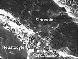
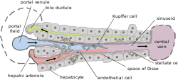
Liver sinusoidal endothelial cells (here LSEC), form the walls of the smallest blood vessels in the liver, also called the hepatic sinusoids. LSECs are highly specialized endothelial cells with characteristic morphology. The LSECs are an important part of the reticuloendothelial system.
Structure
[edit]The LSECs contain many small pores, or fenestrae, with approximate diameters of 100 to 150 nm, which provide open channels between the sinusoidal blood and the subendothelial space of Disse[1]. The fenestrae lack a diaphragm and are clustered together in groups, referred to as “sieve plates”. The fenestrae are crucial for lipoprotein traffic between the hepatocytes and blood [2]. Chylomicrons produced by the intestinal epithelial cells from dietary lipids have diameter up to 1000 nm which makes them unable to pass through the fenestrae. Chylomicrons are metabolized by lipoprotein lipase on endothelial cells of systemic capillaries. The chylomicron remnants are smaller particles (30‐80 nm) and pass through the LSEC fenestrations, to be metabolised in hepatocytes.
Function
[edit]LSECs play an important role in clearance of blood borne waste, immunology, liver regeneration, and liver fibrosis. The clearance of blood-borne waste was previously attributed to Kupffer cells (KC). However, in recent decades it has become clear that LSECs and KCs play a complementary role in this process. This is the concept of the dual cell principle of waste clearance; LSECs clear macromolecules and nanoparticles roughly <200 nm by clathrin-mediated endocytosis whereas KCs clear larger particles >200 nm by phagocytosis [3]. LSECs express some receptors normally found on cells of the immune system and play a role in immunity by scavenging pathogens <200 nm. The LSECs express a number of endocytosis receptors that mediate extremely rapid internalization of waste molecules. In rat it has been shown that they express scavenger receptors class A, B, E and H. The latter is devided into stabilin-1 (SR-H1) and stabilin-2 (SR-H2) being the most important. LSECs also express high levels of the mannose receptor and the Fc-gamma receptor IIb2. Other important receptors on LSECs are L-SIGN (liver/lymph node-specific ICAM-3 grabbing nonintegrin), LSECtin (liver and lymph node sinusoidal endothelial cell C-type lectin), Lyve-1 (lymphatic vessel endothelial hyaluronan receptor‐1), and LRP‐1 (low‐density lipoprotein receptor‐related protein‐1)[4]
History
[edit]The LSEC ultrastructure was first described in detail by Wisse in 1970 [5]. Wisse's detailed electron microscopy study of perfusion fixed rat liver and described the LSEC as a unique cell type, clearly different from the KC. In the eighties, the physiological significance of LSECs as scavenger cells was established [6] when it was found that the cells represented the major site of clearance of blood‐borne connective tissue molecules like hyaluronan, chondroitin sulfate, heparin, N-terminal propeptides of procollagen types I and III, serglycin, nidogen, collagen -chains (types I, II, II, IV, V and XI). Later, in the nineties, the role of LSECs in immunity was found. The cells express a number of pattern recognition receptors (PPRs) like cells of the innate immune system. Furthermore it has been reported that LSECs play a significant role in liver immune tolerance [7].
See also
[edit]References
[edit]- ^ Braet, F; Wisse, E (23 August 2002). "Structural and functional aspects of liver sinusoidal endothelial cell fenestrae: a review". Comparative hepatology. 1 (1): 1. PMID 12437787.
- ^ Fraser, R; Dobbs, BR; Rogers, GW (March 1995). "Lipoproteins and the liver sieve: the role of the fenestrated sinusoidal endothelium in lipoprotein metabolism, atherosclerosis, and cirrhosis". Hepatology (Baltimore, Md.). 21 (3): 863–74. PMID 7875685.
- ^ Sørensen, KK; McCourt, P; Berg, T; Crossley, C; Le Couteur, D; Wake, K; Smedsrød, B (15 December 2012). "The scavenger endothelial cell: a new player in homeostasis and immunity". American journal of physiology. Regulatory, integrative and comparative physiology. 303 (12): R1217-30. doi:10.1152/ajpregu.00686.2011. PMID 23076875.
- ^ Sørensen, KK; Simon-Santamaria, J; McCuskey, RS; Smedsrød, B (20 September 2015). "Liver Sinusoidal Endothelial Cells". Comprehensive Physiology. 5 (4): 1751–74. doi:10.1002/cphy.c140078. PMID 26426467.
- ^ Wisse, E (April 1970). "An electron microscopic study of the fenestrated endothelial lining of rat liver sinusoids". Journal of ultrastructure research. 31 (1): 125–50. PMID 5442603.
- ^ Smedsrød, B; Pertoft, H; Gustafson, S; Laurent, TC (1 March 1990). "Scavenger functions of the liver endothelial cell". The Biochemical journal. 266 (2): 313–27. doi:10.1042/bj2660313. PMID 2156492.
{{cite journal}}: More than one of|author1=and|last1=specified (help) - ^ Knolle, PA; Wohlleber, D (May 2016). "Immunological functions of liver sinusoidal endothelial cells". Cellular & molecular immunology. 13 (3): 347–53. doi:10.1038/cmi.2016.5. PMID 27041636.
