User:Wall277/sandbox
Description
[edit]
Engrams or memory traces are a hypothetical means by which memory traces are stored as biophysical or biochemical changes in the brain (and other neural tissue) in response to external stimuli.
They are sometimes thought of as a neural network or fragment of memory, sometimes using a hologram analogy to describe its action, in light of results showing that memory appears not to be localized in the brain. The existence of engrams is posited by some scientific theories to explain the persistence of memory and how memories are stored in the brain. The existence of neurologically defined engrams is not significantly disputed, though their exact mechanism and location has been a focus of persistent research for many decades.
Overview
[edit]The term engram was coined by the little-known but influential memory researcher Richard Semon.
The engram is a physical change in a brain region that is associated with learning [1].
At the beginning of the twentieth century, Shepherd Ivory Franz attemped to solve the problem [2]. He believed that learning and retention was found in the frontal lobes, but after lesion reseach in cats and monkeys the findings indicated otherwise [2]. Franz with the knowledge of his research and brain-damaged humans came to the conclusion, that the brain has anatomical distinct regions that are not associated with specialized functions [2].
Franz and Lashley teamed up and carried out two experiments on learning [2]. The second experiment examined the effects of lesions in cortical regions in rats by a one-choice maze and an inclined-plane problem [2]. Cortical damage did not affect the maze habit, but frontal lobe damaged affected retention of the inclined-plane problem [2]. After these findings Lashley introduced the term equipotentiality, concluding that at least some part of the frontal lobe must be intact to preserve retention [2].
Karl S. Lashley's search for the engram found that it could not exist in any specific part of the rat's brain, but that memory was widely distributed throughout the cortex [2]. Lashley stated that the entire cortex is required for learning and retention of a complex maze [2]. It did not matter where the lesions were, but rather how large the lesions were as they produced proportional decrement in performance [2]. This indicated that all parts of the brain are active in memory storage, but no one part is more important than another, a principle called equipotentiality [2]. This principle suggests that copies of one memory are spread throughout many brain regions that are responsible for memory [1]. Another term coined by Lashley is mass-action, impairments in learning and retention are proportional to the amount of cortical damage [2]. This term was later disproved experimentally, but the search continues [3]. Lashley also tested brightness discrimination habits and found that they were most likely to be disrupted by lesions in the occipital cortex [4].
One possible explanation for Lashley's failure to locate the engram is that many types of memory (e.g. visual-spatial, smell, etc.) are used in the processing of complex tasks, such as rats running mazes. The consensus view in neuroscience is that the sorts of memory involved in complex tasks are likely to be distributed among a variety of neural systems, yet certain types of knowledge may be processed and contained in specific regions of the brain.[5]. Overall, the mechanisms of memory are poorly understood. Such brain parts as the left parietal cortex, prefrontal regions, striatum, hippocampus, entorhinal cortex and the amygdala are thought to play an important role in memory [1]. For example, the hippocampus is believed to be involved in spatial and declarative learning, as well as consolidating short-term into long-term memory. Also, Lashely may have chosen a memory task (learning of a maze) that was too complex, as it requires many brain regions; as well as his choice of examined brain region, as other regions than the cortex may be involved in memory [6].
Since Lashley's work there has been progress in the search for the engram [6]. Neuron activity has been detected through cellular imaging and is correlated with memory retention, these active neurons may possibly make up memory traces [6].
Experiments
[edit]In Lashley's experiments (1929, 1950), rats were trained to run a maze. Tissue was removed from their cerebral cortices before re-introducing them to the maze, to see how their memory was affected. Increasingly, the amount of tissue removed degraded memory, but more remarkably, where the tissue was removed from made no difference.[5]. Lashley also trained monkeys to do different tasks and later introduced lesions to their brains [1]. After a lesion was introduced the monkey’s memory was tested [1]. The idea was that if you destroy the area of the brain that held the memory of the task (engram), the monkey would not be able to perform the task [1]. Upon experimentation it was found that, the more brain tissue damaged the more time was needed to relearn the task [1]. None of the monkeys actually forgot how to perform the task [1] as did none of the rats.
Later, Richard F. Thompson sought the engram in the cerebellum, rather than the cerebral cortex. He used classical conditioning of the eyelid response in rabbits in search of the engram. He puffed air upon the cornea of the eye and paired it with a tone. (This puff normally causes an automatic blinking response. After a number of experiences associating it with a tone, the rabbits became conditioned to blink when they heard the tone even without a puff.) The experiment monitored several brain regions, trying to locate the engram.
One region that Thompson's group studied was the lateral interpositus nucleus (LIP). When it was deactivated chemically, the rabbits lost the conditioning; when re-activated, they responded again, demonstrating that the LIP is a key element of the engram for this response.[7].
This approach, targeting the cerebellum, though successful, examines only basic, automatic responses, which almost all animals possess, especially as defense mechanisms.
Studies have shown that declarative memories move between the limbic system, deep within the brain, and the outer, cortical regions. These are distinct from the mechanisms of the more primitive cerebellum, which dominates in the blinking response and receives the input of auditory information directly. It does not need to "reach out" to other brain structures for assistance in forming some memories of simple association.
Removal of the superior temporal gyrus or the medial temporal lobe impaired monkeys after the training of an auditory recognition memory task [8].
In 2006 it was found that modality-specific aspects of concepts are differentially located within the brain [9]. Four modalities were tested, including vision, sound, touch and taste by feature verification in an fMRI [2]. Therefore, brain regions that were activated for taste items were not activated for the other three modalities or the semantic control trials [9]. From this study it was concluded that different brain regions correspond to different modalities of knowledge [9]. Therefore, a concept has a distributed pattern of activation across the brain [9]. The problem then arises of how all these modality specific aspects of a concept come together and combine. It has been found that the features of a concept are bound together in the anterior temporal pole [10]. Therefore, all the information of a concept feeds into this area and binds together to create the concept of apple. An apple is red, round, stem on top, tastes good, fruit, grown on trees, etc.
Learning
[edit]Learning occurs when an event alters the nervous system resulting in a change in behaviour, this altered nervous system is a resulting memory [1]. Memory is thought to create a neural circuit upon storage and can be reactivated in the future to experience the memory [1]. Evolutionary the nervous system allows organisms to remember the past to adapt to novel situations [1].
Synaptic plasticity is the cellular level of memory and it is the capacity for change in a synapse between neurons [1]. Donald Hebb took this principle and created the Hebb rule that states, when two neurons that are connected and are both activated the synapse between them is strengthened [1]. The neural change resulting from this Hebb rule is called long-term potentiation (LTP) [1]. LTP will result in increased receptors and increased neurotransmitter release following rapid repeated stimulation of the synapse [1]. Therefore, when one neuron fires the the other neuron will most likely fire as well, due to their strong connection. LTP has been extensively studied in the cells of the hippocampal system [1].

Hippocampal System
[edit]Consolidation is the process by which information stabilizes over time in long-term memory[11]. This term coined by Ebbinhaus, as scientific investigation led him to believe that memory traces were formed gradually over time [12]. Consolidation refers to two types of processes: synaptic consolidation, which occurs within minutes to hours after learning; and system consolidation, which takes much longer and is dependent on the hippocampus for reorganization[11]. Recently, it has been debated that there is a form of renewed consolidation where memories must undergo consolidation each time they are activated as compared to memories only being consolidated once. [11].
There is still no answer as to how memories consolidate, but a possible explanation may be a new protein that is synthesized for memory formation and storage [13]. Further research is required to stabilize this 40 year debate.
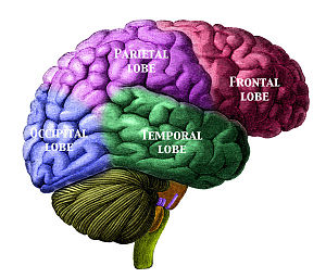
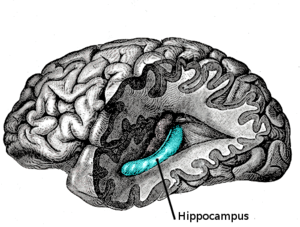
The hippocampus when damaged may lead to anterograde amnesia, which is the inability to remember any new information after the damage [1]. Anterograde amnesia affected the patient H.M. which is discussed later [1]. Retrograde amnesia is the ability to remember information that was before the event that caused the damage [1]. The hippocampus and associated areas is responsible for consolidation [1]. The hippocampus takes records of current events and stores them in a more permanent state [1]. When an organism is experiencing an event, sensory information is pouring into all the brain lobes and needs to be organized and combined. The hippocampus takes all the parts of an experience and combines them to have a single memory [1]. The brains lobes are illustrated in the following picture.

Case Study: H.M.
[edit]H.M. suffered from epilepsy, a disorder which causes spasms and seizures from uncontrollable neuron stimulation [1]. Anticonvulsant medication was ineffective, so surgeons decided to remove portions of his medial temporal lobes, including his hippocampus [1]. After surgery H.M. no longer suffered from seizures but instead had severe memory loss [1]. H.M. could no longer learn any new information [1]. Consolidation could no longer occur as the medial temporal lobes were required for the process of transferring information from working memory to long-term memory [1].
H.M. still had many other memory abilities like working memory and problem-solving abilities [1]. Hig long-term memory for both declaractive and procedural knowledge, prior to the surgery was also unaffected [1]. H.M. was a big contributor to the understanding of memory. This case shows that memory is not in only one area in the brain, but distributed. H.M.`s memory functions are found in different brain areas [1].
Working Memory
[edit]Working memory can be divided into three main areas [1]. The articulatory loop that manages verbal representations; the visuo-spatial sketchpad that processes visual information; and the executive control system that coordinates actions [1]. There is now neuropsychological evidence that confirms that these structures are distinct and found in different areas of the brain [1].
Verbal material is stored in the left hemisphere's posterior parietal cortex [1]. The rehearsal of verbal material is found in three distinct areas of the prefrontal cortex one being the inferior frontal gyrus (Broca's area) [1]. Therefore, for the left hemisphere's posterior parietal cortex and the prefrontal cortex are the brain regions required for the articulatory loop, that is part of the working memory model [1]. Spatial information in working memory was found to be stored in the right hemisphere's posterior parietal cortex [1]. The maintenance of spatial information was found to be in the dorsolateral prefrontal cortex [1]. Finally, a third working memory system was found for objects [1]. This system remembers visual object representations, whereas spatial information only refers to the location of the object, not its appearance [1]. These two systems result in two different pathways for the "where" and the "what" of an object [1]. Spatial memory requires occipital, parietal, and frontal sites in the right hemisphere, whereas object memory requires parietal sites in the left hemisphere [1]. Therefore, one can conclude that there are many distinct systems required for working memory that correspond to different sensory modalities.
Long-Term Memory
[edit]When sensory information arrives at the brain it is scattered in many areas of the cortex, depending on the sensory modality. Without the hippocampal system the representations would quickly fade away. The hippocampus takes the sensory information from the cortex, integrates the information into a whole and then returns the consolidated memory to the cortex [1]. Therefore, the cortex is where long-term memories are stored. Long-term memory comes in different forms. Procedural memories stores how we do things, the skill knowledge and are demonstrated through action [1]. Procedural memory does not require conscious attention unlike declarative memories. Declarative memories store facts and general world knowledge, which are separated into semantic memory for facts and episodic memory for events [1]. Research has confirmed that there are different brain areas that correspond to different types of long-term memory [1].
Declaractive memories are stored in the cortex due to the hippocampus consolidation. It has been suggested that the hippocampus is used for consolidation for episodic memory, but the limbic system controls semantic memory [1]. Therefore if both the hippocampus and the limbic system were destroyed, declarative memory would be lost [1]. Procedural memory also does not depend on the hippocampus for consolidation [1]. The basal ganglia is critical for skill learning, with a minor role for the motor cortex [1].
Category:Memory Category:Neuropsychology
</ref>
- References
- ^ a b c d e f g h i j k l m n o p q r s t u v w x y z aa ab ac ad ae af ag ah ai aj ak al am an ao ap aq ar as at au av aw ax Friedenberg, Jay (2012). Cognitive Science An Introduction to the Study of Mind. California: SAGE Publications, Inc. pp. 171-184. ISBN 978-1-4129-7761-6
- ^ a b c d e f g h i j k l m ^ Bruce, Darryl (2001). "Fifty Years Since Lashley's In Search of the Engram:". Journal of the History of the Neurosciences 10 (3): 308-318.
- ^ ^Hübener, Mark; Bonhoeffer, Tobias (2010). "Searching for Engrams". Neuron. 67 (3): 363–371. doi:10.1016/j.neuron.2010.06.033. PMID 20696375. Retrieved 13 March 2012.
{{cite journal}}: Unknown parameter|month=ignored (help)CS1 maint: date and year (link) - ^ ^Lashley, KS (1920). Studies of cerebral function in learning. Psychobiology 2: 55±135.
- ^ a b Gerrig and Zimbardo (2005) Psychology and Life (17th edition: International edition)
- ^ a b c ^Josselyn, Sheena A. (April 2010). "Continuing the search for the engram: examining the". J Psychiatry Neurosci 35 (4): 221-228. doi:10.1503/jpn.100015.
- ^ James W. Kalat, Biological Psychology p. 392–393
- ^ ^Fritz, Jonathan; Mishkin, Mortimer; Saunders, Richard C. (2005). "In search of an auditory engram". PNAS. 102 (26): 9359–9364. doi:10.1073/pnas.0503998102. JSTOR 3375900. PMC 1166637. PMID 15967995. Retrieved 13 March 2012.
{{cite journal}}: Unknown parameter|month=ignored (help)CS1 maint: date and year (link) - ^ a b c d ^Goldberg, R.F., Perfetti, C.A., Schneider, W. (2006). Perceptual knowledge retrieval activates sensory brain regions. Journal of Neuroscience, 26, 4917-4921
- ^ ^Patterson K, Nestor PJ, Rogers TT (2007), “Where do you know what you know? The representation of semantic knowledge in the human brain.” Nat Rev Neurosci 8(12):976-87
- ^ a b c Dudai, Yadin (2004). "The Neurobiology of Consolidations,or,How Stable is the Engram?". Annu. Rev. Psychol. 55: 51–86. doi:10.1146/annurev.psych.55.090902.142050. PMID 14744210. Retrieved 13 March 2012.
- ^ Sara, Susan J.; Hars, Bernard (2006). "In memory of consolidation". Learning & Memory. 13 (5): 515–521. doi:10.1101/lm.338406. PMID 17015848. Retrieved 13 March 2012.
{{cite journal}}: CS1 maint: date and year (link) - ^ Klann, Eric; Sweatt, J. David (2008). "Altered protein synthesis is a trigger for long-term memory formation". Neurobiology of Learning and Memory. 89 (3): 247–259. doi:10.1016/j.nlm.2007.08.009. PMC 2323606. PMID 17919940. Retrieved 13 March 2012.
{{cite journal}}: Check date values in:|year=/|date=mismatch (help); Unknown parameter|month=ignored (help)

