User:Terrasigillata/Apicalectodermalridge
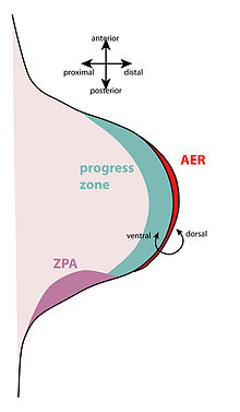
The apical ectodermal ridge (AER) is a thickened layer of ectodermal cells at the distal tip of a developing limb bud. Along with the zone of polarizing activity, it is a crucial organizing region during limb development.

Morphology and Role
[edit]The AER consists of tightly packed columnar cells at the dorso-ventral boundary on a growing limb bud. Continued signaling from the AER to the progress zone, the layer of mesenchyme immediately proximal to the AER, induces cell growth, migration, and differentiation during limb development.
The position of the limb bud, and hence the AER, is specified by the expression boundaries of Hox genes in the embryonic trunk. At these positions, the induction of cell outgrowth is thought to be mediated by a positive feedback loop of fibroblast growth factors between the intermediate mesoderm, the lateral plate mesoderm and the surface ectoderm. FGF-8 in the intermediate mesoderm signals to the lateral mesoderm, restricting the expression of FGF-10 through intermediate Wnt signals. Then, FGF-10 in the lateral plate mesoderm signals to the surface ectoderm to create the AER, which expresses FGF8[Ref 1].
The AER is known to express FGF-2, FGF-4, FGF-8, and FGF-9, while the limb bud mesenchyme expresses FGF-2 and FGF-10. Embryo manipulation experiments have shown that some of these FGFs alone are sufficient for mimicking the AER[Ref 2].
Transplantation experiments
[edit]Removal/Addition of AER
[edit]The removal of the AER results in truncated limbs where only the stylopod is present [Ref 3]. The transplantation of an additional AER results in the duplication of limb structures, usually as a mirror image next to the already developing limb. The mirror image reflection is a result of the transplanted AER obeying signals from the existing ZPA.
FGF soaked beads can mimic the AER
[edit]Implantation of a plastic bead soaked in FGF-4 or FGF-2 will induce formation of a limb bud in an embryo, but proliferation will cease prematurely unless additional beads are added to maintain appropriate levels of the FGF. Implantation of sufficient beads can induce formation of a 'normal' additional limb at an arbitrary location in the embryo [Ref 4] [Ref 5].
Transplantation of the AER to flank mesoderm between the normal limb buds results in ectopic limbs. If the AER is transplanted closer to the forelimb bud, the ectopic limb develops like a forelimb. If the AER is transplanted closer to the hindlimb bud, the ectopic limb develops like a hindlimb[Ref 6]. If the AER is transplanted near the middle, the ectopic limb has both forelimb and hindlimb features[Ref 7].
AER does not specify limb identity
[edit]Transplantation of an AER that would give rise to an arm (or wing, as these experiments are commonly performed on chicken embryos) to a limb field developing into a leg does not produce an arm and leg at the same location, but rather two legs. In contrast, transplantation of cells from the progress zone of a developing arm to replace the progress zone of a developing leg will produce a limb with leg structures proximally (femur, knee) and arm structures distally (hand, fingers). Thus it is the mesodermal cells of the progress zone, not the ectodermal cells of the AER, that control the identity of the limb [Ref 8].
AER timing does not specify underlying mesoderm fate
[edit]AER timing does not regulate the fate specification of the underlying mesoderm, as shown by one set of experiments (cite here). When the AER from a late limb bud is transplanted to an earlier limb bud, the limb forms normally. The converse – transplantation of an early limb bud to a late limb bud – also results in normal limb development. However, the underlying mesoderm in the progress zone ‘’is’’ fate specified. If progress zone mesoderm is transplanted along with the AER, then additional finger/toes are formed (for an early-->late transplantation) or the finger/toes are formed too early (for a late-->early transplantation)[Ref 3].
External links
[edit]- http://embryology.med.unsw.edu.au/Notes/skmus7a.htm
- http://www.ncbi.nlm.nih.gov/bookshelf/br.fcgi?book=dbio&part=A3941#A3954
References
[edit]- ^ Ohuchi, H., Nakagawa, T., Yamamoto, a., Araga, a., Ohata, T., Ishimaru, Y., et al. (1997). The mesenchymal factor, FGF10, initiates and maintains the outgrowth of the chick limb bud through interaction with FGF8, an apical ectodermal factor. Development (Cambridge, England), 124(11), 2235-44. Retrieved from http://www.ncbi.nlm.nih.gov/pubmed/9187149.
- ^ Martin, G. R. (1998). The roles of FGFs in the early development of vertebrate limbs. Genes & Development, 12(11), 1571-1586. doi: 10.1101/gad.12.11.1571.
- ^ a b Rubin, L., & Saunders, J. W. (1972). Ectodermal-Mesodermal in the Chick Embryo : Interactions Constancy Ectodermal in the Growth and Temporal of Limb Buds Limits of the Induction ’. Developmental Biology, 112, 94-112.
- ^ Fallon, J. F., López, a., Ros, M. a., Savage, M. P., Olwin, B. B., Simandl, B. K., et al. (1994). FGF-2: apical ectodermal ridge growth signal for chick limb development. Science (New York, N.Y.), 264(5155), 104-7. Retrieved from http://www.ncbi.nlm.nih.gov/pubmed/7908145.
- ^ Niswander, L., Tickle, C., Vogel, a., Booth, I., & Martin, G. R. (1993). FGF-4 replaces the apical ectodermal ridge and directs outgrowth and patterning of the limb. Cell, 75(3), 579-87. Retrieved from http://www.ncbi.nlm.nih.gov/pubmed/8221896.
- ^ Cohn, M. J., Izpisúa-Belmonte, J. C., Abud, H., Heath, J. K., & Tickle, C. (1995). Fibroblast growth factors induce additional limb development from the flank of chick embryos. Cell, 80, 739-746.
- ^ Ohuchi, H., Takeuchi, J., Yoshioka, H., Yoshiyasu, I., Ogura, K., Takahashi, N., et al. (1998). Correlation of wing-leg identity in ectopic FGF-induced chimeric limbs with the differential expression of chick Tbx5 and Tbx4.
- ^ Zwilling, E. (1959). Interaction between ectoderm and mesoderm in duck–chicken limb bud chimaeras. Journal of Experimental Zoology, 142, 521–532.
