User:Talkingneanderthal/ACF sandbox
| Anterior cranial fossa | |
|---|---|
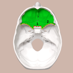
 Superior view of the skull base. Anterior cranial fossa shown in green. 1: Sphenoidal limbus (anterior margin of the chiasmatic groove) | |
 Base of skull. Superior view. Boundaries of the cranial fossae drawn as red lines. | |
| Details | |
| Identifiers | |
| Latin | fossa cranii anterior |
| Anatomical terminology | |
The anterior cranial fossa is a depression in the floor of the cranial base which houses the projecting frontal lobes of the brain. It is formed by the horizontally-oriented orbital plates of the frontal bone, the cribriform plate of the ethmoid bone, and the lesser wings (orbitosphenoids) and body of the sphenoid bone. This part of the sphenoid body can be referred to as the jugum sphenoidale, planum sphenoidale, or presphenoid bone, depending on context. The anterior margin of the chiasmatic groove is situated directly posterior to the jugum sphenoidale and runs on either side to the upper margin of the optic foramen. The posterior margin of the anterior cranial fossa is at the junction between the lesser wings and anterior margin of the chiasmatic groove laterally and the tuberculum sellae along the midline and just to either side. The anterior cranial fossa is positioned anterior to the middle cranial fossa, and the posterior, middle, and anterior cranial fossae are arranged like a staircase when viewed from the side, with the anterior cranial fossa occupying the "highest" position closest to the vertex of the crania.
Anatomical Description
[edit]The frontal, ethmoid, and sphenoid bones contact each another in the anterior cranial fossa at the frontoethmoidal, sphenoethmoidal, and sphenofrontal sutures. In humans, the sphenoid and ethmoid bones typically contact each other broadly across the midline, the contact involving both the presphenoid and lesser wings, although in a smaller number of cases the two bones contact each other via a sphenoid or ethmoid process or spine. In a small number of cases the orbital plates of frontal bone intervene between the sphenoid and ethmoid bones to meet in the midline at a "retro-ethmoid" frontal suture.
The lateral portions of the anterior cranial fossa form the roof over each orbit and the platform on which the frontal lobes of the cerebrum rest. The orbital plates of the anterior cranial fossa are convex and marked by depressions and grooves that more or less faithfully mark the locations of brain convolutions and branches of meningeal blood vessels.
The midline portion of the anterior cranial fossa is both the roof of the nasal cavity and the bony platform that supports the olfactory bulbs. In humans, the horizontal plate of ethmoid bone (called the cribriform plate) is riddled with small holes that transmit olfactory nerves, and the nasociliary nerve runs through a slit-like opening between the ethmoid and frontal bones at the cribriform plate's anterior margin. The cribriform plate forms an olfactory groove on each side of the midline. Two midline structures - the root of the frontal crest and the crista galli - are attachment points for the dural falx cerebri, and the crista galli separates the right and left olfactory bulbs. The foramen cecum, which is located between the frontal crest and crista galli, usually transmits a small vein from the nasal cavity to the superior sagittal sinus.
Openings
[edit]A number of openings on the floor of the anterior cranial fossa transmit nerves and blood vessels between the endocranium, nasal cavity, and orbits. The internal openings of the anterior and posterior ethmoidal foramina are situated lateral to each olfactory groove. The anterior openings, which are situated halfway along the lateral margin of the olfactory groove, transmit the anterior ethmoidal vessels and nasociliary nerve, the latter of which runs in a groove along the lateral edge of the cribriform plate to the slit-like opening at its anterior margin. The posterior ethmoidal foramen, which opens at the posterior margin under cover of the projecting lamina of the sphenoid, transmits the posterior ethmoidal vessels and nerve. The anterior margin of the chiasmatic groove is situated directly posterior to the @ and runs on either side to the upper margin of the optic foramen, through which the optic nerve runs.
Contents
[edit]The anterior cranial fossa contains the following parts of the brain:
- frontal lobes of the cerebral cortex,
- olfactory bulbs,
- olfactory tracts,
- orbital gyri.[1]
Openings
[edit]There are several openings connecting the anterior cranial fossa with other parts of the skull, including the:
- anterior ethmoidal foramen,
- cribriform foramina.
The paired anterior ethmoidal foramen connects the anterior cranial fossa with each orbit and transmits the anterior ethmoidal artery, nerve and vein.
The cribriform foramina are the openings in the cribriform plate of the ethmoid bone, which connect the anterior cranial fossa with the nasal cavity and transmit the olfactory nerves.[2]
- ^ "Anterior cranial fossa". www.anatomynext.com. Retrieved 2018-03-06.
- ^ "Anterior cranial fossa". www.anatomynext.com. Retrieved 2018-03-06.
