User:Sransom2/sandbox
Temporoparietal Junction
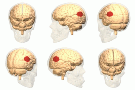
The temporoparietal junction is a component of the human brain, located in between the parietal lobe and temporal lobe. It is defined as the area where these two major lobes meet. The temporoparietal junction consists of two main lobes: left temporoparietal junction and right temporoparietal junction. As a result of brain lateralization, the left and right lobes function independently of each other. Damage to the temporoparietal junction most often results in out of body experiences (OBE). Lesion studies have shown the right temporoparietal junction plays a major role in the regulation of attention, metacognition, and spatial awareness. Lesion studies in the left temporoparietal junction show a correlation to inference about others beliefs or theory of the mind and speech perception.
Anatomy
[edit]


The temporoparietal junction is a region in the brain located between the parietal lobe and temporal lobe. It is positioned near the posterior end of the lateral sulcus, a fissure which divides the hemispheres of the parietal lobe, temporal lobe, and frontal lobe. The main functions of the parietal lobe are attention and spatial processing [1] . It has also been known to govern cognition, speech, and visual perception [1].The main functions of the temporal lobe include: auditory processing, pattern recognition, and language comprehension [1].The temporoparietal junction lies in the centre of the brain, separated by the cerebral fissure. The ventral stream of the brain, the 'what' pathway responsible for object recognition, also flows through this area [1]. The 'what' pathway is responsible for object recognition. The temporoparietal junction can be divided into two parts: the left lobe which is located in the left hemisphere, and the right lobe which is located in the right hemisphere. Because the lobes are located in different hemispheres, they are said to be lateralized and therefore, differ in function. The left hemisphere of the brain specializes in logical, analytical, and grammatical thinking. This area has poor spatial resolution. The right hemisphere is known for artistic and concrete thinking, with excellent spatial orientation [2]
Theory of the Mind
[edit]The Theory of Mind (TOM) proposes that humans are able to relate mental states to both the self and to others in order to understand behaviour[3]. This includes beliefs, intents, knowledge, and ideas. Essentially, it involves 'mind-reading' by allowing an individual to understand why a person is acting a certain way and how they will likely act in the future [4]. A particular region of the temporoparietal junction, TPJ-M, functions by using the TOM to understand the intentions of others [3]. Damage to the temporoparietal junction shows deficits in the ability to utilize TOM. Theory of the Mind is also absent in individuals who have autism, schizophrenia, and ADHD [5].
Theory of the Mind is not directly observable; it is simply a quality of the mind [6]. A problem that can arise from this is the mind-body problem which questions the compatibility between the physical brain and the mental mind. The body and the brain are material, whereas the mind is subjective. Aristotle proposed the theory of Monism which states the mind and brain are a single entity: either completely physical (known as physicalism) or completely mental (known as idealism)[1] . Plato introduced another theory, known as Dualism, proposing that the brain and the mind are two separate entities: the mind is mental and the brain is physical [1]. To this day, there is still debates as to which theory is correct. For more information on this debate, refer to the book Aristotle and Plotinus on the Intellect: Monism and Dualism Revisited by Mark J. Nyvlt.
Aside from understanding the beliefs of others, TOM also attempts to understand the underlying thoughts of the self [1] . This is done through the process of introspection, or 'looking inward' to reflect on one's own conscious thoughts [1] . It is essentially the ability to think about thinking. Introspection allows humans to relate to others by developing a thorough understanding of their own thought processes.
A study by Saxe and Kanwisher, used fMRI testing to measure blood oxygen level dependence (BOLD) in response to given tasks[3]. Subjects were asked to read two stories: one that described characters mental states and another that focused on the physical qualities of the characters, relating them closely to non-human objects. A higher BOLD signal was measured in the TPJ-M of subjects who read stories about mental states compared to those who read stories about physical qualities [3]. This study further supports the belief that TOM is controlled by the temporoparietal junction.
Lesions
[edit]
The easiest way to study the function of a part of the brain is to impair that region while keeping the surrounding areas intact. After impairing the area of interest, many techniques are used to study the repercussions, including: behaviour observation, magnetic resonance imaging (MRI), and positron emission tomography (PET). There are two ways to study brain impairments: animal induced lesion studies and human case studies [1]. In animal studies, brain damage can be induced on the area of interest and animal behaviour can then be studied to note any changes as a result of damage [1]. In humans, it would be unethical to induce lesions and therefore we must instead look at case studies -- patients who have accidentally acquired brain damage in the given area [1]. These may occur as a result of many things, including head trauma, chemical mutagens, stroke, or seizure. It is important to keep in mind that case studies are not very reliable because patients often differ on many other variables (i.e. race, origin, gender, development, etc), therefore it is difficult to determine whether the observed functioning is a result of the damage or one of the other variables [1]. Case studies do however provide us with insight which could not otherwise be studied.
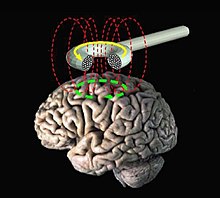
One ethical way to study lesions in humans would be through the use of transcranial magnetic stimulation (TMS). In TMS, a large magnetic coil is placed over the patient's head, emitting an electrical current which causes neurons in the brain to fire [1]. This current can either be used to stimulate neurons or cause temporary lesions. These lesions are harmless and can be very useful in studying behaviour. After the lesion has been created, the patient is observed while performing a given task and the behavioural changes are noted [1].
Lesion studies have been used to examine the functions of the temporoparietal junction. Lesion studies of this area have tended to focus on human patients, using both TMS and case studies. Lesions in the temporoparietal junction are specific to the right lobe and left lobe. These studies are discussed in great detail in the "Functions" sections for each lobe shown below.
Left Temporoparietal Junction
[edit]The left temporoparietal junction is the part of the brain where the left temporal lobe and the left parietal lobe meet. Research about the function of the left temporoparietal junction is very limited. Few studies have been able to draw conclusive evidence about this area of the brain, perhaps because it is very difficult to stimulate such a small precise area without also stimulating the lobes around it.
Although we may not know everything about the function of the left temporoparietal junction, there are some areas that it does play a role in. A large field of research that the left temporoparietal lobe has been linked to is Theory of the Mind and inference about the beliefs of other people. Studies have suggested that the left temporoparietal junction is singly responsible for the proper functioning of TOM. Another area of interest is speech perception and the regulatory role the left temporoparietal lobe plays in mediating the pathway between the primary motor cortex and posterior inferior frontal gyrus.
Inference About Others Beliefs
[edit]Neuroscientists have found the major role of the left temporoparietal lobe to be involved in inference about the beliefs of others. This is known as Theory of the Mind (TOM), described in detail a section above. The ability to infer about the beliefs of others is distributed across the various areas of the brain: the prefrontal cortex, posterior cingulate, and the left temporoparietal lobe [7] .
Samson and colleagues studied TOM in the left temporoparietal lobe by examining 3 case studies[8] . Sampson tested the three patients on various mental state reasoning tasks: language, executive functioning, and gaze and pointing tasks. He compared these results to a control group (a group of individuals without damage to the left temporoparietal junction). Sampson determined the individuals with damage did significantly worse on these tasks [8]. This research supports the notion that the left temporoparietal junction mediates inference about the beliefs of others.
Speech Perception
[edit]Speech perception, the comprehension of spoken language, is localized in auditory cortex of the temporal lobe. It is the ability to hear and understand auditory input. Auditory information flows through the sensorimotor cortex after passing through the temporal lobe [9]. Language enters the brain in the form of phonemes (the smallest unit of sound) and is understood at the pragmatics level (rules of conversation) [1].
Murakami, a neurologist at Goethe University, conducted a study to test the links between speech perception and the left temporoparietal lobe. Murakami induced lesions in healthy patients using transcranial magnetic stimulation (TMS) to study the flow of information through the primary motor cortex (M1) and the left temporoparietal lobe [9]. Upon stimulation of the left temporoparietal junction, patients could no longer comprehend speech [9] . Studying further, Murakami found that the TMS also created lesions in the posterior inferior frontal gyrus (pIFG) and M1. He concluded that the flow of information must flow from the left temporoparietal junction, through M1, into pIFG. Without a fully functional left temporoparietal lobe, information cannot pass through this pathway [9] . This supports the notion that the left temporoparietal junction is necessary for speech perception.
Right Temporoparietal Junction
[edit]The right parietal junction is the area where the right temporal lobe and right parietal lobe meet. There have been numerous studies done on the right temporoparietal junction, including both case studies and lesion studies. Many studies have found conclusive evidence regarding the functions of the right temporoparietal junction. As this area has been found to have many specializations, it supports the theory of distributed coding -- meaning that many different areas of the brain are responsible for cohesively working together to control a single function[1]. This provides evidence against modularity of mind -- a theory explaining how the brain is divided into separate modules, each of which performs a specific function [1].
The right temporoparietal junction is responsible for many functions, including: attention, metacognition, and spatial unity of the body. Through the use of transcranial magnetic stimulation (TMS) [10] and brain imaging from magnetic resonance imaging [11], the right temporoparietal lobe has been directly linked to attention. Studies have also shown the right temporoparietal lobe to be connected to bottom-up mental processing [12]. Perhaps the most influential area that the right temporoparietal lobe is known for is spatial unity. Damage in this area causes out of body experiences (OBE), a condition in which patients feel separated from their body. Many case studies have examined epileptic patients who suffer from OBE's with damage specifically to the right temporoparietal junction. Please note that many of the functions of the right temporoparietal junction can be directly related to the functions of the temporal lobe and parietal lobe (described in detail above).
Attention
[edit]Attention can be described as the mind's ability to concentrate [1] . Attention can be classified into two categories: selective (focused on one source at a time) and divided (focused on several sources at a time) [1] . Because attention is important in many aspects of brain functioning, it is known to be distributed across many areas of the brain, including but not limited to: the reticular activating system (RAS), superior colliculus, thalamus, intraparietal sulcus, cingulate cortex, frontal lobe, and the temporoparietal junction [1]. Without cohesion between all of these brain areas, attention would not be able to function properly.
Research has shown a strong correlation between the temporoparietal junction and salience (i.e. pain) and attention [11]. Previous studies have used magnetic resonance imaging to visualize the brains of healthy patients in comparison to patients with temporoparietal lobe damage [11]. Results have shown that a stronger connectivity lies in the right temporoparietal lobe rather than the left parietal lobe [11].
Meister, a neuroscientist from University of Toronto, attempted to explain the connection between the superior temporal gyrus (STG) and the right temporoparietal lobe [10]. He used a visual-spatial attention task, along with transcranial magnetic stimulation (TMS) to study this relationship [10] . Lesions created using TMS in the STG showed no effect on completion of attention tasks. However, temporoparietal lesions showed a decrease in attentional capacity [10], indicating that information flows first through the temporoparietal junction rather than through the STG. He concluded the temporoparietal junction is responsible for visual attention.
Metacognition
[edit]Metacognition is the regulation or control of any aspect of cognition: planning, resource allocation, checking, and error detection [1]. Essentially, it is having an awareness of one's own knowledge. Metacognition was believed to have been located in the prefrontal cortex [1], however, new studies suggest it may be distributed throughout the brain with a connection to the right temporoparietal lobe. Metacognition is an important part of problem-solving: it evaluates the effectiveness of a strategy and considers the usefulness of alternative strategies [1]. Disfunction of metacognition is associated with persistant use of inappropriate problem solving strategies [1].
Decety and Lamm conducted a meta-analysis of 70 studies involving neuroimaging [12]. Findings suggest that the right temporoparietal lobe mediates bottom-up mental processing [12], meaning it uses information from the environment to form an idea. The study proposes that social cognition is domain-general, meaning the parts of the brain work together in cohesion, rather than each having specific functions.[12].
Body Awareness and Spatial Unity: Out of Body Experiences
[edit]
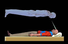
An out of body experience (OBE) can be thought of as a dissociation between the mind and the physical body; a feeling of being separated and seeing the body and surroundings from an 'outside' perspective [13][14]. Studies have directly linked OBE's to damage in the right temporoparietal lobe [15]. Out of body experiences do not simply affect certain areas of the brain, rather it effects the entire body as a whole [13], inducing a sense of fear and disconnection. During the experience of an OBE, the individual appears to be awake and aware of what is occurring [14]. Patients have reported feelings of floating while experiencing an OBE [14]. There is currently no testable neuroscientific study to measure OBE's [15] aside from observing case studies.
Olaf Blanke and colleagues studied 6 OBE patients using nueroimaging-- a test that produces a visual image of the brain, allowing researchers to observe functioning. Blanke discovered a few notable symptoms of OBE patients: vestibular sensations (i.e. floating or flying), body‐part illusions, and seeing the body as impartial (not whole)[15]. Through brain imaging, researchers determined 5 of the 6 patients with OBE's had damage to the temporoparietal junction [15], implying that the temporoparietal lobe mediates spatial unity.
A study by Genevan neurologists, Heydrich and colleagues, focused on a case study of a 10-year-old female who suffered from focal epilepsy [13]. One of the main symptoms the patient experienced was OBE's which occurred simultaneously with epileptic seizures [13]. Through the use of Magnetic resonance imaging (MRI), Electroencephalography (EEG), and Positron Emission Tomography (PET), the researches discovered lesions in the girl's right temporoparietal junction [13]. This suggests that the OBE's are a direct result of damage to the right temporoparietal area. A study by Blanke and colleagues also studied an epileptic patient and found similar results [14].
Other research has examined the effects of transcranial magnetic stimulation (TMS) on the right temporoparietal area of healthy patients [14]. One study asked healthy patients to imagine being disconnected from the body (replicating OBE sensations) and then used event related potential (ERP) to measure brain activity [14]. When asked to perform the task, patients showed activation in the right temporoparietal junction [14]. They performed a second study which looked at brain images of patients in the absence of TMS (deemed the control group) compared to brain images of patients with TMS targeted at the right temporoparietal junction (deemed the treatment group)[14]. Researchers concluded that individuals with lesions in the right temporoparietal region induced by TMS had lost the ability to imagine spatial movements of objects and/or the body in space [14], supporting the notion that the temporoparietal junction has spatial localization.
Summary and Future Research
[edit]The temporoparietal lobe has separate functions for the left and right lobe. The left lobe is majorly responsible for inference about others beliefs and speech perception. Aside from these functions, it is still unknown what this area is for. In the future, this area should be studied in more depth. Lesions should be induced using transcranial magnetic stimulation (TMS). By asking patients to carry out tasks during TMS, we can study the impaired regions. In addition, another way to study this area would be through case studies. Unfortunately, because case studies require accidental damage, it may take some time before a case study with damage in this region presents itself. The right temporoparietal junction is specifically involved in attention, metacognition, and out of body experiences. Although a decent amount of research has been conducted on the right temporoparietal junction, it would be beneficial to explore this region further. Studies have shown that this area may also be involved in musical ability. Future research should look into this concept. All in all, there is still a lot to be discovered about the temporoparietal junction.
See Also
[edit]References
[edit]- ^ a b c d e f g h i j k l m n o p q r s t u v w x Friedenberg, J., & Silverman, G. (2012). Cognitive Science (2nd ed.) United States of America: SAGE Publications, Inc.
- ^ Meerwaldt, J. D., & Van Harskamp, F. (1982). Spatial disorientation in right-hemisphere infarction. Journal of Neurology, Neurosurgery & Psychiatry, 45(7), 586-590.
- ^ a b c d Saxe, R., & Kanwisher, N. (2005). People thinking about thinking people: The role of the temporo-parietal junction in "theory of mind". (pp. 171-182). New York, NY, US: Psychology Press, New York, NY.
- ^ Robbins, P. (2004). Knowing me, knowing you: Theory of mind and the machinery of introspection. Journal of Consciousness Studies, 11(7-8), 129-143.
- ^ Korkmaz B (May 2011). "Theory of mind and neurodevelopmental disorders of childhood". Pediatr. Res. 69 (5 Pt 2): 101R–8R.
- ^ David Premack and Guy Woodruff (1978). Does the chimpanzee have a theory of mind?. Behavioral and Brain Sciences, 1, pp 515-526.
- ^ Saxe, R. (2006). Four brain regions for one theory of mind? (pp. 83-101). Cambridge, MA, US: MIT Press, Cambridge, MA.
- ^ a b Samson, D., Apperly, I. A., Chiavarino, C., & Humphreys, G. W. (2004). Left temporoparietal junction is necessary for representing someone else’s belief. Nature Publishing Group, 7(5), 499-500.
- ^ a b c d Murakami, T., Restle, J., & Ziemann, U. (2012). Effective connectivity hierarchically links temporoparietal and frontal areas of the auditory dorsal stream with the motor cortex lip area during speech perception. Brain and Language, 122(3), 135-141.
- ^ a b c d Meister, I. G., Weinemann, M., Buelte, D., Grunewald, C., Sparing, R., Dambeck, N., & Boroojerdi, B. (2006). Hemiextinction induced by transcranial magnetic stimulation over the right temporo-parietal junction. Neuroscience, 142, 119-123.
- ^ a b c d Kucyi, A., Hodaie, M., & Davis, K. D. (2012). Lateralization in intrinsic functional connectivity of the temporoparietal junction with salience- and attention-related brain networks. Journal of Neurophysiology, 108(12), 3382-3392.
- ^ a b c d Decety, J., & Lamm, C. (2007). The role of the right temporoparietal junction in social interaction: How low-level computational processes contribute to meta-cognition. The Neuroscientist, 13(6), 580-593.
- ^ a b c d e Heydrich, L., Lopez, C., Seeck, M., & Blanke, O. (2011). Partial and full own-body illusions of epileptic origin in a child with right temporoparietal epilepsy. Epilepsy & Behavior, 20(3), 583-586.
- ^ a b c d e f g h i Blanke, O., Mohr, C., Michel, C. M., Pascual-Leone, A., Brugger, P., Seeck, M., Thut, G. (2005). Linking out-of-body experience and self processing to mental own-body imagery at the temporoparietal junction. The Journal of Neuroscience, 25(3), 550-557.
- ^ a b c d Blanke, O., Landis, T., Spinelli, L., & Seeck, M. (2004). Out-of-body experience and autoscopy of neurological origin. Brain: A Journal of Neurology, 127(2), 243-258.

