User:Snake playing a saxaphone/histology of nerve entrapment
Histology [deprecated?]
[edit]Nerve entrapment is treated with a decompression rather than a biopsy, so human tissue of nerve entrapment is rare.[1]
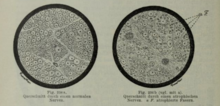
The predominant finding is a thinning of myelin in myelinated fibers. This is due to the end stages of demyelination or the early stages of remyelination.[1]
The degree of demyelination can vary within a fascicle and between fascicles.[1][2]
The histopathological findings in the scarce human tissue samples are consistent with the results found in animal models.[2]
The histopathology changes most in the area under compression.[2]
Increased microvascular permeability is an early indicator of tissue injury. For nerve compression this is the step in the pathophysiology cascade and is detected with the use of Evans blue albumin. This substance is injected before nerve tissue harvested and the spread of this blue dye allows the visual tracking of vascular permeability.[3]
Renaut bodies are seen in histology of compressed nerves although the function of Renaut bodies remains unclear.[4]
[You can keep this if you talk about histological changes seen like myelin / axon ratio]
[The thinning myelin image would be nice]
Histological studies are the foundation of assessing structural changes caused by nerve entrapment.
[Mention that data from animal studies is in agreement with human tissue samples]

Histological studies of compressed nerve show abnormalities in the compressed region.[2]
Animal models are where the bulk of nerve pathophysiology research has been done. The two most common animal models are rabbits and rats. Studies have been done acute nerve compression[3], chronic nerve compression[5], nerve stretch[6], nerve adhesions[7], repetitive compression[8], etc.
Human studies are the best model for nerve entrapment but suffers from lack of availability of nerve tissue.[5] Nerve biopsies aren't generally used to study nerve entrapment because a biopsy will result in permanent nerve dysfunction[9]. Access to human tissue generally comes from cadavers[1] or a sensory nerve like the sural nerve, LFCN, ACN when the nerve is resected anyway as a medical treatment. Due to the poor availability of human nerve tissue, it is not possible to study a progression of nerve entrapment but rather the available tissue acts as a snapshot at that specific moment in the disease progression that can be correlated to animal models.
Human nerve tissue under various stages of compression is generally limited to case studies. Examples of some cases studies are the sural nerve, radial nerve, anterior cutaneous abdominal nerve, and lateral femoral cutaneous nerve.
[Human studies are hard because biopsies will mean a Sunderland 5 injury]
[this section is probably important to show what the findings of resected nerves are]
[meralgia paresthetica]
In a study on resected LFCN nerves, the major findings where: multi-focal fiber loss, reduced fiber density, selective loss of large myelinated fibers, perineurial thickening, subperineurial edema, and renaut bodies. These features were not seen in the control nerves.[10]
[sural nerve biopsy]
[radial nerve][2]
[anterior cutaneous abdominal nerve]
In a study on ACNES, resected anteriour cutaneous abdominal nerves were examined histopathologically for evidence of inflammation or infection as an alternative cause. No evidence of either of those disease process was found in any patients.[11]
- ^ a b c d Mackinnon SE. Pathophysiology of nerve compression. Hand Clin. 2002 May;18(2):231-41. doi: 10.1016/s0749-0712(01)00012-9. PMID: 12371026.
- ^ a b c d e Mackinnon SE, Dellon AL, Hudson AR, Hunter DA. Chronic human nerve compression--a histological assessment. Neuropathol Appl Neurobiol. 1986 Nov-Dec;12(6):547-65. doi: 10.1111/j.1365-2990.1986.tb00159.x. PMID: 3561691.
- ^ a b Rydevik B, Lundborg G. Permeability of intraneural microvessels and perineurium following acute, graded experimental nerve compression. Scand J Plast Reconstr Surg. 1977;11(3):179-87. doi: 10.3109/02844317709025516. PMID: 609900.
- ^ Skidmore RA, Woosley JT, Tomsick RS. Renaut bodies. Benign disease process mimicking neurotropic tumor infiltration. Dermatol Surg. 1996 Nov;22(11):969-71. PMID: 9063513.
- ^ a b O'Brien JP, Mackinnon SE, MacLean AR, Hudson AR, Dellon AL, Hunter DA. A model of chronic nerve compression in the rat. Ann Plast Surg. 1987 Nov;19(5):430-5. doi: 10.1097/00000637-198711000-00008. PMID: 3688790.
- ^ Wall EJ, Massie JB, Kwan MK, Rydevik BL, Myers RR, Garfin SR. Experimental stretch neuropathy. Changes in nerve conduction under tension. J Bone Joint Surg Br. 1992 Jan;74(1):126-9. doi: 10.1302/0301-620X.74B1.1732240. PMID: 1732240.
- ^ Abe Y, Doi K, Kawai S. An experimental model of peripheral nerve adhesion in rabbits. Br J Plast Surg. 2005 Jun;58(4):533-40. doi: 10.1016/j.bjps.2004.05.012. PMID: 15897039.
- ^ Yoshii Y, Nishiura Y, Terui N, Hara Y, Saijilafu, Ochiai N. The effects of repetitive compression on nerve conduction and blood flow in the rabbit sciatic nerve. J Hand Surg Eur Vol. 2010 May;35(4):269-78. doi: 10.1177/1753193408090107. Epub 2009 Aug 17. PMID: 20444785.
- ^ National Research Council (US) Steering Committee for the Workshop on Work-Related Musculoskeletal Injuries: The Research Base. Work-Related Musculoskeletal Disorders: Report, Workshop Summary, and Workshop Papers. Washington (DC): National Academies Press (US); 1999. Biological Response of Peripheral Nerves to Loading: Pathophysiology of Nerve Compression Syndromes and Vibration Induced Neuropathy. Available from: https://www.ncbi.nlm.nih.gov/books/NBK230871/
- ^ Berini SE, Spinner RJ, Jentoft ME, Engelstad JK, Staff NP, Suanprasert N, Dyck PJ, Klein CJ. Chronic meralgia paresthetica and neurectomy: a clinical pathologic study. Neurology. 2014 Apr 29;82(17):1551-5. doi: 10.1212/WNL.0000000000000367. Epub 2014 Mar 28. PMID: 24682967; PMCID: PMC4011467.
- ^ Markus J, van Montfoort M, de Jong JR, de Beer SA, Aronica EMA, Gorter RR. Histopathologic examination of resected nerves from children with anterior cutaneous nerve entrapment syndrome: Clues for pathogenesis? J Pediatr Surg. 2020 Dec;55(12):2783-2786. doi: 10.1016/j.jpedsurg.2020.01.060. Epub 2020 Feb 22. PMID: 32156426.
