User:CFCF/Grant
Appearance
-
Grant 1962 3 -
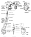
Superficial Veins of the Upper Limb
(Dissected and drawn by Miss Nancy Joy.)
The arrows indicate where perforating veins pierce the deep fascia and bring the superficial and deep veins of the limb into communication with each other.
For obvious mechanical reasons the palmar veins are few and small, and the dorsal veins are large, as seen in figures 5 and 6. -
Superficial Veins of the Hand - Palmar Aspect
(Dissected and drawn by Miss Nancy Joy.)
See legend attached to figure 4. -
Superficial Veins of the Hand - Dorsal Aspect
-
Cutaneous Nerves of the Upper Limb
Of the 5 terminal branches of the brachial plexus, viz. musculo-cutaneous, medain, ulnar radial, and axillary (circumflex) nerves, the first 4 reach the hand.
The posterior cord of the plexus is represented by 5 cutaneous nerves. Of these (a) one, the upper lateral cutaneous n. of the arm, is a branch of the axillary n.;
(b) whereas 4 are branches of the radial n. They are: the posterior cutaneous n. of the arm, the lower lateral cutaneous n. of the arm, the posterior cutaneous n. of the forearm, and the superficial branch of the radial nerve.
See figures 13 (pectoral region), 24 (back), 44 (elbow), 83 (hand). -
Cutaneous Nerves of the Upper Limb
See 7 -
Scheme of the Motor Distribution of the Ventral Nerves of the Limb
The average levels at which the motor branches leave the stems of the main nerves are shown with reference to the lower border of the axilla (Teres Major), elbow joint (medical epicondyle), and wrist (pisiform bone). -
Scheme of the Motor Distribution of the Dorsal Nerves of the Limb
The average levels of origin of the motor branches are shown as in figure 9.


