User:Bob K31416/CRC
Pathology
[edit]The pathology of the tumor is usually reported from the analysis of tissue taken from a biopsy or surgery. A pathology report will usually contain a description of cell type and grade. The most common colon cancer cell type is adenocarcinoma which accounts for 95% of cases. Other, rarer types include lymphoma and squamous cell carcinoma.
Cancers on the right side (ascending colon and cecum) tend to be exophytic, that is, the tumour grows outwards from one location in the bowel wall. This very rarely causes obstruction of feces, and presents with symptoms such as anemia. Left-sided tumours tend to be circumferential, and can obstruct the bowel much like a napkin ring.
Adenocarcinoma is a malignant epithelial tumor, originating from glandular epithelium of the colorectal mucosa. It invades the wall, infiltrating the muscularis mucosae, the submucosa and thence the muscularis propria. Tumor cells describe irregular tubular structures, harboring pluristratification, multiple lumens, reduced stroma ("back to back" aspect). Sometimes, tumor cells are discohesive and secrete mucus, which invades the interstitium producing large pools of mucus/colloid (optically "empty" spaces) - mucinous (colloid) adenocarcinoma, poorly differentiated. If the mucus remains inside the tumor cell, it pushes the nucleus at the periphery - "signet-ring cell." Depending on glandular architecture, cellular pleomorphism, and mucosecretion of the predominant pattern, adenocarcinoma may present three degrees of differentiation: well, moderately, and poorly differentiated.[1]
Most colorectal cancer tumors are thought to be cyclooxygenase-2 (COX-2) positive. This enzyme is generally not found in healthy colon tissue, but is thought to fuel abnormal cell growth.
-
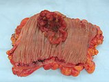
Appearance of the inside of the colon showing one invasive colorectal carcinoma (the crater-like, reddish, irregularly shaped tumor).
-
Gross appearance of a colectomy specimen containing two adenomatous polyps (the brownish oval tumors above the labels, attached to the normal beige lining by a stalk) and one invasive colorectal carcinoma (the crater-like, reddish, irregularly shaped tumor located above the label).
-
Endoscopic image of colon cancer identified in sigmoid colon on screening colonoscopy in the setting of Crohn's disease.
-
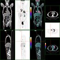
PET/CT of a staging exam of colon carcinoma. Besides the primary tumor a lot of lesions can be seen. On cursor position: lung nodule.
-
Cancer — Invasive adenocarcinoma (the most common type of colorectal cancer). The cancerous cells are seen in the center and at the bottom right of the image (blue). Near normal colon-lining cells are seen at the top right of the image.
-
Cancer — Histopathologic image of colonic carcinoid.
-
Precancer — Tubular adenoma (left of image), a type of colonic polyp and a precursor of colorectal cancer. Normal colorectal mucosa is seen on the right.
-
Precancer — Colorectal villous adenoma.
For colorectal cancer patients in the US, 42% have diagnosed malignancies in the right side of the colon, which consists of the cecum, ascending colon, hepatic flexure, and transverse colon.[2]
According to the International Classification of Diseases for Oncology, for the purpose of describing tumor location as right-sided or left-sided, the right side of the colorectum consists of the cecum, ascending colon, hepatic flexure, and transverse colon, and the left side consists of the splenic flexure, descending colon, sigmoid colon, recto-sigmoid junction, and rectum.[3][4]
- ^ Pathology atlas
- ^ "Trends in Colorectal Cancer Incidence Rates in the United States by Tumor Location and Stage, 1992–2008". Cancer Epidemiology, Biomarkers & Prevention. 21 (3). 411–6 (See Introduction). Mar 2012. doi:10.1158/1055-9965.EPI-11-1020. PMID 22219318. Retrieved 2012-09-16.
{{cite journal}}: Unknown parameter|authors=ignored (help) - ^ "Trends in Colorectal Cancer Incidence Rates in the United States by Tumor Location and Stage, 1992–2008". Cancer Epidemiology, Biomarkers & Prevention. 21 (3). 411–6 (See section Methods — Statistical analysis). Mar 2012. doi:10.1158/1055-9965.EPI-11-1020. PMID 22219318. Retrieved 2012-09-16.
{{cite journal}}: Unknown parameter|authors=ignored (help) - ^ International Classification of Diseases for Oncology (ICD-O) (Third ed.). World Health Organization. 1 Jan 2000. ISBN 978-9241545341.
{{cite book}}: Unknown parameter|authors=ignored (help)
- Trends in Colorectal Cancer Incidence by Anatomic Site and Disease Stage in the United States From 1976 to 2005 — PMID: 21217399


