User:BarneyStinson13/sandbox
Ankle Sprain
[edit]Anatomy
[edit]The ankle is a ginglymus joint made up of a series of bones and ligaments.

This joint allows for the dorsiflexion and plantar flexion of the foot. The ankle is composed of the lateral malleolus, inferior tibiofibular joint, medial malleolus, talus and calcaneus. The primary ligaments of the ankle include the inferior deltoid ligament, the anterior/posterior inferior tibofibular ligaments, anterior/posterior talofibular ligaments, calcaneofibular ligament, calcaneofibular ligament, interosseus ligament, interosseus membrane, and the Achilles tendon.
Mechanism
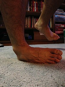
[edit][Ankle sprains] occur usually through excessive stress on the ligaments of the ankle. This is can be caused by excessive external rotation, inversion or eversion of the foot caused by an external force. When the foot is moved past its range of motion, the excess stress puts a strain on the ligaments. If the strain is great enough to the ligaments past the yield point, then the ligament becomes damaged, or sprained[1][2]
Injury
[edit]
When the ankle becomes inverted, the anterior talofibular and calcaneofibular ligaments are damaged. This is the most common ankle sprain. Another common ankle sprain is caused by external rotation of the foot[3]. This will strain the inferior deltoid ligament, and in extreme cases, the Achilles tendon.

References
[edit]- ^ Wikstrom, Erik A., Wikstrom, April M., Hubbard-Turner, Tricia., Ankle Sprains: Treating to Prevent the Long-Term Consequences. JAAPA. October 2012. 25(10)
- ^ Gehring, D., Wissler, S., Mornieux, G., Gollhofer, A., How to Sprain Your Ankle – A Biomechanical Case Report of an Inversion Trauma. Journal of Biomechanics. 46 (2013) 175-178
- ^ Norkus, Susan A., Floyd, R.T., The Anatomy and Mechanisms of Syndesmotic Ankle Sprains. Journal of Athletic Training. 2001; 36(1):68-73
