Sindbis virus
| Sindbis virus | |
|---|---|

| |
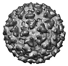
| CryoEM model of Sindbis virus. EMDB entry EMD-2374[1] | |
| Virus classification | |
| (unranked): | Virus |
| Realm: | Riboviria |
| Kingdom: | Orthornavirae |
| Phylum: | Kitrinoviricota |
| Class: | Alsuviricetes |
| Order: | Martellivirales |
| Family: | Togaviridae |
| Genus: | Alphavirus |
| Species: | Sindbis virus
|
Sindbis virus (SINV) is a member of the Togaviridae family, in the Alphavirus genus. The virus was first isolated in 1952 in Cairo, Egypt.[2] The virus is transmitted by mosquitoes (Culex and Culiseta). SINV is linked to Pogosta disease[3] (Finland), Ockelbo disease (Sweden) and Karelian fever (Russia). In humans, the symptoms include arthralgia, rash and malaise. Sindbis virus is widely and continuously found in insects and vertebrates in Eurasia, Africa, and Oceania. Clinical infection and disease in humans however has almost only been reported from Northern Europe (Finland, Sweden, Russian Karelia), where SINV is endemic and where large outbreaks occur intermittently. Cases are occasionally reported in Australia, China, and South Africa.[4]
SINV is an arbovirus, it is arthropod-borne, and it is maintained in nature by transmission between vertebrate (bird) hosts and invertebrate (mosquito) vectors. Humans are infected with Sindbis virus when bitten by an infected mosquito.
Virus physiology
[edit]Structure, genome & replication
[edit]Sindbis viruses are enveloped particles with an icosahedral capsid, with a positive single stranded RNA genome, with an approximate size of 11.7 kb. The RNA has a 5'-cap and 3'-polyadenylated tail, and therefore serves directly as messenger RNA (mRNA) in a host cell. The genome encodes four non-structural proteins, the capsid, and two envelope proteins. This is characteristic of all Togaviruses. Replication is cytoplasmic and rapid. The genomic RNA is partially translated at the 5’ end to produce the non-structural proteins which are then involved in genome replication and the production of new genomic RNA and a shorter sub-genomic RNA strand. This sub-genomic strand is translated into the structural proteins. The viruses assemble at the host cell surfaces and acquire their envelope through budding.
A non-coding RNA element has been found to be essential for Sindbis virus genome replication.[5]
Recombination has been demonstrated between RNAs of Sindbis virus.[6][7] The mechanism of recombination appears to be template switching (copy choice) during RNA replication.[6][7]
See also
[edit]References
[edit]- MicrobiologyBytes: Togaviruses
- CDC: Pogosta disease and Sindbis virus
- Sindbis virus—ICTVdB—The Universal Virus Database, version 4.
- ^ Cao, S.; Zhang, W. (2013). "Characterization of an early-stage fusion intermediate of Sindbis virus using cryoelectron microscopy". Proceedings of the National Academy of Sciences. 110 (33): 13362–13367. Bibcode:2013PNAS..11013362C. doi:10.1073/pnas.1301911110. PMC 3746934. PMID 23898184.
- ^ Ling J, Smura T, Lundström JO, Pettersson JH, Sironen T, Vapalahti O, Lundkvist Å, Hesson JC (30 July 2019). "Introduction and Dispersal of Sindbis Virus from Central Africa to Europe". J Virol. 93 (16): e00620-19. doi:10.1128/JVI.00620-19. PMC 6675900. PMID 31142666.
- ^ Kurkela S, Manni T, Vaheri A, Vapalahti O. Causative agent of Pogosta disease isolated from blood and skin lesions, Emerg Infect Dis [serial on the Internet]. Published 2004 May. (accessed 2007-10-16)
- ^ "Facts about Sindbis fever". European Centre for Disease Prevention and Control. Retrieved 7 September 2021.
- ^ Frolov, I; Hardy R; Rice CM (2001). "Cis-acting RNA elements at the 5' end of Sindbis virus genome RNA regulate minus- and plus-strand RNA synthesis". RNA. 7 (11): 1638–1651. doi:10.1017/S135583820101010X. PMC 1370205. PMID 11720292.
- ^ a b Lai MM (1992). "RNA recombination in animal and plant viruses". Microbiol Rev. 56 (1): 61–79. doi:10.1128/mr.56.1.61-79.1992. PMC 372854. PMID 1579113.
- ^ a b Weiss BG, Schlesinger S (1991). "Recombination between Sindbis virus RNAs". J Virol. 65 (8): 4017–25. doi:10.1128/JVI.65.8.4017-4025.1991. PMC 248832. PMID 2072444.
