SUCLG2
| SUCLG2 | |||||||||||||||||||||||||||||||||||||||||||||||||||
|---|---|---|---|---|---|---|---|---|---|---|---|---|---|---|---|---|---|---|---|---|---|---|---|---|---|---|---|---|---|---|---|---|---|---|---|---|---|---|---|---|---|---|---|---|---|---|---|---|---|---|---|
| Identifiers | |||||||||||||||||||||||||||||||||||||||||||||||||||
| Aliases | SUCLG2, GBETA, succinate-CoA ligase GDP-forming beta subunit, G-SCS, GTPSCS, succinate-CoA ligase GDP-forming subunit beta | ||||||||||||||||||||||||||||||||||||||||||||||||||
| External IDs | OMIM: 603922; MGI: 1306824; HomoloGene: 2854; GeneCards: SUCLG2; OMA:SUCLG2 - orthologs | ||||||||||||||||||||||||||||||||||||||||||||||||||
| |||||||||||||||||||||||||||||||||||||||||||||||||||
| |||||||||||||||||||||||||||||||||||||||||||||||||||
| |||||||||||||||||||||||||||||||||||||||||||||||||||
| |||||||||||||||||||||||||||||||||||||||||||||||||||
| |||||||||||||||||||||||||||||||||||||||||||||||||||
| Wikidata | |||||||||||||||||||||||||||||||||||||||||||||||||||
| |||||||||||||||||||||||||||||||||||||||||||||||||||
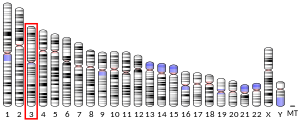
Succinyl-CoA ligase [GDP-forming] subunit beta, mitochondrial is an enzyme that in humans is encoded by the SUCLG2 gene on chromosome 3.[5]
This gene encodes a GTP-specific beta subunit of succinyl-CoA synthetase. Succinyl-CoA synthetase catalyzes the reversible reaction involving the formation of succinyl-CoA and succinate. Alternate splicing results in multiple transcript variants. Pseudogenes of this gene are found on chromosomes 5 and 12. [provided by RefSeq, Apr 2010][5]
Structure
[edit]SCS, also known as succinyl CoA ligase (SUCL), is a heterodimer composed of a catalytic α subunit encoded by the SUCLG1 gene and a β subunit encoded by either the SUCLA2 gene or the SUCLG2 gene, which determines the enzyme specificity for either ADP or GDP. SUCLG2 is the SCS variant containing the SUCLG2-encoded β subunit.[6][7][8] Amino acid sequence alignment of the two β subunit types reveals a homology of ~50% identity, with specific regions conserved throughout the sequences.[9]
SUCLG2 is located on chromosome 3 and contains 14 exons.[5]
Function
[edit]As a subunit of SCS, SUCLG2 is a mitochondrial matrix enzyme that catalyzes the reversible conversion of succinyl-CoA to succinate and acetoacetyl CoA, accompanied by the substrate-level phosphorylation of GDP to GTP, as a step in the tricarboxylic acid (TCA) cycle.[6][7][8][10] The GTP generated is then consumed in anabolic pathways.[7][9] However, since GTP is not transported through the inner mitochondrial membrane in mammals and other higher organisms, it must be recycled within the matrix.[8] In addition, SUCLG2 may function in ATP generation in the absence of SUCLA2 by complexing with the mitochondrial nucleotide diphosphate kinase, nm23-H4, and thus compensate for SUCLA2 deficiency.[6][8] The reverse reaction generates succinyl-CoA from succinate to fuel ketone body and heme synthesis.[6][8]
While SCS is ubiquitously expressed, SUCLG2 is predominantly expressed in tissues involved in biosynthesis, including liver and kidney.[8][9][11] SUCLG2 has also been detected in the microvasculature of the brain, likely to support its growth.[7] Notably, both SUCLA2 and SUCLG2 are absent in astrocytes, microglia, and oligodendrocytes in the brain; thus, in order to acquire succinate to continue the TCA cycle, these cells may instead synthesize succinate through GABA metabolism of α-ketoglutarate or ketone body metabolism of succinyl-CoA.[7][8]
Clinical significance
[edit]Though mitochondrial DNA (mtDNA) depletion syndrome has been largely attributed to SUCLA2 deficiency, SUCLG2 may play a more crucial role in mtDNA maintenance, as it functions to compensate for SUCLA2 deficiency and its absence results in decreased mtDNA and OXPHOS-dependent growth.[6] Moreover, no mutations in the SUCLG2 gene have been reported, indicating that such mutations are lethal and selected against.[8]
SUCLG2 may also play a role in clearing cerebrospinal fluid amyloid-beta 1–42 (Aβ1–42) in Alzheimer's disease (AD) and, thus, reducing neuronal death.[10]
See also
[edit]References
[edit]- ^ a b c GRCh38: Ensembl release 89: ENSG00000172340 – Ensembl, May 2017
- ^ a b c GRCm38: Ensembl release 89: ENSMUSG00000061838 – Ensembl, May 2017
- ^ "Human PubMed Reference:". National Center for Biotechnology Information, U.S. National Library of Medicine.
- ^ "Mouse PubMed Reference:". National Center for Biotechnology Information, U.S. National Library of Medicine.
- ^ a b c "Entrez Gene: SUCLA2 succinate-CoA ligase, GDP-forming, beta subunit".
- ^ a b c d e Miller C, Wang L, Ostergaard E, Dan P, Saada A (May 2011). "The interplay between SUCLA2, SUCLG2, and mitochondrial DNA depletion" (PDF). Biochimica et Biophysica Acta (BBA) - Molecular Basis of Disease. 1812 (5): 625–9. doi:10.1016/j.bbadis.2011.01.013. PMID 21295139.
- ^ a b c d e Dobolyi A, Bagó AG, Gál A, Molnár MJ, Palkovits M, Adam-Vizi V, Chinopoulos C (April 2015). "Localization of SUCLA2 and SUCLG2 subunits of succinyl CoA ligase within the cerebral cortex suggests the absence of matrix substrate-level phosphorylation in glial cells of the human brain" (PDF). Journal of Bioenergetics and Biomembranes. 47 (1–2): 33–41. doi:10.1007/s10863-014-9586-4. PMID 25370487. S2CID 41101828.
- ^ a b c d e f g h Dobolyi A, Ostergaard E, Bagó AG, Dóczi T, Palkovits M, Gál A, Molnár MJ, Adam-Vizi V, Chinopoulos C (January 2015). "Exclusive neuronal expression of SUCLA2 in the human brain" (PDF). Brain Structure & Function. 220 (1): 135–51. doi:10.1007/s00429-013-0643-2. PMID 24085565. S2CID 105582.
- ^ a b c Johnson JD, Mehus JG, Tews K, Milavetz BI, Lambeth DO (October 1998). "Genetic evidence for the expression of ATP- and GTP-specific succinyl-CoA synthetases in multicellular eucaryotes". The Journal of Biological Chemistry. 273 (42): 27580–6. doi:10.1074/jbc.273.42.27580. PMID 9765291.
- ^ a b Ramirez A, van der Flier WM, Herold C, Ramonet D, Heilmann S, Lewczuk P, Popp J, Lacour A, Drichel D, Louwersheimer E, Kummer MP, Cruchaga C, Hoffmann P, Teunissen C, Holstege H, Kornhuber J, Peters O, Naj AC, Chouraki V, Bellenguez C, Gerrish A, Heun R, Frölich L, Hüll M, Buscemi L, Herms S, Kölsch H, Scheltens P, Breteler MM, Rüther E, Wiltfang J, Goate A, Jessen F, Maier W, Heneka MT, Becker T, Nöthen MM (December 2014). "SUCLG2 identified as both a determinator of CSF Aβ1-42 levels and an attenuator of cognitive decline in Alzheimer's disease". Human Molecular Genetics. 23 (24): 6644–58. doi:10.1093/hmg/ddu372. PMC 4240204. PMID 25027320.
- ^ Matilainen S, Isohanni P, Euro L, Lönnqvist T, Pihko H, Kivelä T, Knuutila S, Suomalainen A (March 2015). "Mitochondrial encephalomyopathy and retinoblastoma explained by compound heterozygosity of SUCLA2 point mutation and 13q14 deletion". European Journal of Human Genetics. 23 (3): 325–30. doi:10.1038/ejhg.2014.128. PMC 4326715. PMID 24986829.


