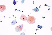
Navicular cell
Appearance

Navicular cell is a boat-shaped benign epithelial cell seen in Pap smear.[1] They are seen in pregnancy (most prominently during smears taken in the second trimester),[2] second half of menstrual cycle, during menopause and in women using medroxyprogesterone acetate (depo-provera) for contraception. Navicular cells have folded edges, with a thickened outer rim of cytoplasm and an eccentric nucleus. They contain abundant glycogen in the cytoplasm, giving it a central yellow halo. The cytoplasm appears golden, refractile and granular under the microscope. In depo-provera users, the high progesterone levels result in more exfoliation of superficial squamous cells, thereby causing navicular cells to appear in Pap smear.[3]
References
[edit]- ^ Harshmohan (2010). Harshmohan's textbook of Pathology (5 ed.). New Delhi: Jaypee Publications. p. 280. ASIN 9351523691.
- ^ Sattenspiel, Edward (1950). "Vaginal cytology in normal pregnancy". Obstetrical & Gynecological Survey. 5 (6): 800. doi:10.1097/00006254-195012000-00003. Retrieved 9 December 2016.
- ^ EE, Volk (23 September 2000). "Cytologic findings in cervical smears in patients using intramuscular medroxyprogesterone acetate (Depo-provera) for contraception". Diagnostic Cytopathology. 23 (3): 161–4. doi:10.1002/1097-0339(200009)23:3<161::AID-DC4>3.0.CO;2-O. PMID 10945902. S2CID 39180738.
Gallery
[edit]


