HLA-E
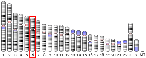
HLA class I histocompatibility antigen, alpha chain E (HLA-E) also known as MHC class I antigen E is a protein that in humans is encoded by the HLA-E gene.[5] The human HLA-E is a non-classical MHC class I molecule that is characterized by a limited polymorphism and a lower cell surface expression than its classical paralogues. The functional homolog in mice is called Qa-1b, officially known as H2-T23.
Structure
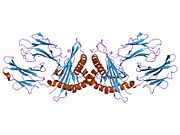
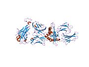
[edit]Like other MHC class I molecules, HLA-E is a heterodimer consisting of an α heavy chain and a light chain (β-2 microglobulin). The heavy chain is approximately 45 kDa and anchored in the membrane. The HLA-E gene contains 8 exons. Exon one encodes the signal peptide, exons 2 and 3 encode the α1 and α2 domains, which both bind the peptide, exon 4 encodes the α3 domain, exon 5 encodes the transmembrane domain, and exons 6 and 7 encode the cytoplasmic tail.[6]
Function
[edit]HLA-E has a very specialized role in cell recognition by natural killer cells (NK cells).[7] HLA-E binds a restricted subset of peptides derived from signal peptides of classical MHC class I molecules, namely HLA-A, B, C, G.[8] These peptides are released from the membrane of the endoplasmic reticulum (ER) by the signal peptide peptidase and trimmed by the cytosolic proteasome.[9][10] Upon transport into the ER lumen by the transporter associated with antigen processing (TAP), these peptides bind to a peptide binding groove on the HLA-E molecule.[11] This allows HLA-E to assemble correctly and to be expressed on the cell surface. NK cells recognize the HLA-E+peptide complex using the heterodimeric receptor CD94/NKG2A/B/C.[7] When CD94/NKG2A or CD94/NKG2B is engaged, it produces an inhibitory effect on the cytotoxic activity of the NK cell to prevent cell lysis. However, binding of HLA-E to CD94/NKG2C (see KLRC2) results in NK cell activation. This interaction has been shown to trigger expansion of NK cell subsets in antiviral responses,[12] where adaptive NK cells that express CD94/NKG2C can specifically recognise HCMV-derived peptide antigens.[13]
References
[edit]- ^ a b c ENSG00000225201, ENSG00000236632, ENSG00000230254, ENSG00000206493, ENSG00000233904, ENSG00000229252 GRCh38: Ensembl release 89: ENSG00000204592, ENSG00000225201, ENSG00000236632, ENSG00000230254, ENSG00000206493, ENSG00000233904, ENSG00000229252 – Ensembl, May 2017
- ^ a b c GRCm38: Ensembl release 89: ENSMUSG00000073405 – Ensembl, May 2017
- ^ "Human PubMed Reference:". National Center for Biotechnology Information, U.S. National Library of Medicine.
- ^ "Mouse PubMed Reference:". National Center for Biotechnology Information, U.S. National Library of Medicine.
- ^ Mizuno S, Trapani JA, Koller BH, Dupont B, Yang SY (Jun 1988). "Isolation and nucleotide sequence of a cDNA clone encoding a novel HLA class I gene". Journal of Immunology. 140 (11): 4024–30. doi:10.4049/jimmunol.140.11.4024. PMID 3131426.
- ^ "Entrez Gene: HLA-E major histocompatibility complex, class I, E".
- ^ a b Braud VM, Allan DS, O'Callaghan CA, Söderström K, D'Andrea A, Ogg GS, Lazetic S, Young NT, Bell JI, Phillips JH, Lanier LL, McMichael AJ (Feb 1998). "HLA-E binds to natural killer cell receptors CD94/NKG2A, B and C". Nature. 391 (6669): 795–9. Bibcode:1998Natur.391..795B. doi:10.1038/35869. PMID 9486650. S2CID 4379457.
- ^ Braud V, Jones EY, McMichael A (May 1997). "The human major histocompatibility complex class Ib molecule HLA-E binds signal sequence-derived peptides with primary anchor residues at positions 2 and 9". European Journal of Immunology. 27 (5): 1164–9. doi:10.1002/eji.1830270517. PMID 9174606. S2CID 36124500.
- ^ Lemberg MK, Bland FA, Weihofen A, Braud VM, Martoglio B (Dec 2001). "Intramembrane proteolysis of signal peptides: an essential step in the generation of HLA-E epitopes". Journal of Immunology. 167 (11): 6441–6. doi:10.4049/jimmunol.167.11.6441. PMID 11714810.
- ^ Bland FA, Lemberg MK, McMichael AJ, Martoglio B, Braud VM (Sep 2003). "Requirement of the proteasome for the trimming of signal peptide-derived epitopes presented by the nonclassical major histocompatibility complex class I molecule HLA-E". The Journal of Biological Chemistry. 278 (36): 33747–52. doi:10.1074/jbc.M305593200. PMID 12821659.
- ^ Braud VM, Allan DS, Wilson D, McMichael AJ (Jan 1998). "TAP- and tapasin-dependent HLA-E surface expression correlates with the binding of an MHC class I leader peptide". Current Biology. 8 (1): 1–10. Bibcode:1998CBio....8....1B. doi:10.1016/S0960-9822(98)70014-4. PMID 9427624. S2CID 14240878.
- ^ Rölle A, Pollmann J, Ewen EM, Le VT, Halenius A, Hengel H, Cerwenka A (Dec 2014). "IL-12-producing monocytes and HLA-E control HCMV-driven NKG2C+ NK cell expansion". The Journal of Clinical Investigation. 124 (12): 5305–16. doi:10.1172/JCI77440. PMC 4348979. PMID 25384219.
- ^ Hammer Q, Rückert T, Borst EM, Dunst J, Haubner A, Durek P, Heinrich F, Gasparoni G, Babic M, Tomic A, Pietra G, Nienen M, Blau IW, Hofmann J, Na IK, Prinz I, Koenecke C, Hemmati P, Babel N, Arnold R, Walter J, Thurley K, Mashreghi MF, Messerle M, Romagnani C (May 2018). "Peptide-specific recognition of human cytomegalovirus strains controls adaptive natural killer cells". Nature Immunology. 19 (5): 453–463. doi:10.1038/s41590-018-0082-6. PMID 29632329. S2CID 4718187.
Further reading
[edit]- Kuby Immunology, 6th edition, by Thomas J. Kindt, Richard A. Goldsby, and Barbara A. Kuby W. H. Freeman and Company, New York
- Moretta L, Bottino C, Pende D, Mingari MC, Biassoni R, Moretta A (May 2002). "Human natural killer cells: their origin, receptors and function". European Journal of Immunology. 32 (5): 1205–11. doi:10.1002/1521-4141(200205)32:5<1205::AID-IMMU1205>3.0.CO;2-Y. PMID 11981807.
- Jensen PE, Sullivan BA, Reed-Loisel LM, Weber DA (Jun 2004). "Qa-1, a nonclassical class I histocompatibility molecule with roles in innate and adaptive immunity". Immunologic Research. 29 (1–3): 81–92. doi:10.1385/IR:29:1-3:081. PMID 15181272. S2CID 29282633.


