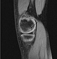
File:OCD WalterReed MRI-Sagital-T1.jpeg
Appearance

Size of this preview: 579 × 600 pixels. Other resolutions: 232 × 240 pixels | 463 × 480 pixels | 945 × 979 pixels.
Original file (945 × 979 pixels, file size: 353 KB, MIME type: image/jpeg)
File history
Click on a date/time to view the file as it appeared at that time.
| Date/Time | Thumbnail | Dimensions | User | Comment | |
|---|---|---|---|---|---|
| current | 21:10, 4 March 2009 |  | 945 × 979 (353 KB) | FoodPuma | Added arrow (edited with Adobe Photoshop CS2) |
| 00:12, 8 February 2009 |  | 516 × 467 (79 KB) | FoodPuma | {{Information |Description={{en|1="Sagittal and coronal T1 and T2 images demonstrate linear low T1, high T2 signal at the articular surfaces of the lateral aspects of the medial femoral condyles bilaterally, corresponding to the radiographs, confirming th |
File usage
The following 2 pages use this file:
Global file usage
The following other wikis use this file:
- Usage on ar.wikipedia.org
- Usage on az.wikipedia.org
- Usage on ca.wikipedia.org
- Usage on he.wikipedia.org
- Usage on ja.wikipedia.org
- Usage on nl.wikipedia.org
- Usage on pl.wikipedia.org
- Usage on uk.wikipedia.org
- Usage on www.wikidata.org
- Usage on zh.wikipedia.org

