Caspase 3
Caspase-3 is a caspase protein that interacts with caspase-8 and caspase-9. It is encoded by the CASP3 gene. CASP3 orthologs[4] have been identified in numerous mammals for which complete genome data are available. Unique orthologs are also present in birds, lizards, lissamphibians, and teleosts.
The CASP3 protein is a member of the cysteine-aspartic acid protease (caspase) family.[5] Sequential activation of caspases plays a central role in the execution-phase of cell apoptosis. Caspases exist as inactive proenzymes that undergo proteolytic processing at conserved aspartic residues to produce two subunits, large and small, that dimerize to form the active enzyme. This protein cleaves and activates caspases 6 and 7; and the protein itself is processed and activated by caspases 8, 9, and 10. It is the predominant caspase involved in the cleavage of amyloid-beta 4A precursor protein, which is associated with neuronal death in Alzheimer's disease.[6] Alternative splicing of this gene results in two transcript variants that encode the same protein.[7]
 |
 |
Caspase-3 shares many of the typical characteristics common to all currently-known caspases. For example, its active site contains a cysteine residue (Cys-163) and histidine residue (His-121) that stabilize the peptide bond cleavage of a protein sequence to the carboxy-terminal side of an aspartic acid when it is part of a particular 4-amino acid sequence.[9][10] This specificity allows caspases to be incredibly selective, with a 20,000-fold preference for aspartic acid over glutamic acid.[11] A key feature of caspases in the cell is that they are present as zymogens, termed procaspases, which are inactive until a biochemical change causes their activation. Each procaspase has an N-terminal large subunit of about 20 kDa followed by a smaller subunit of about 10 kDa, called p20 and p10, respectively.[12]
Substrate specificity
[edit]Under normal circumstances, caspases recognize tetra-peptide sequences on their substrates and hydrolyze peptide bonds after aspartic acid residues. Caspase 3 and caspase 7 share similar substrate specificity by recognizing tetra-peptide motif Asp-x-x-Asp.[13] The C-terminal Asp is absolutely required while variations at other three positions can be tolerated.[14] Caspase substrate specificity has been widely used in caspase based inhibitor and drug design.[15]
Structure
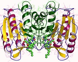
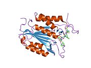
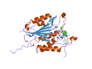
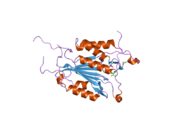
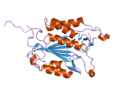
[edit]Caspase-3, in particular, (also known as CPP32/Yama/apopain)[16][17][18] is formed from a 32 kDa zymogen that is cleaved into 17 kDa and 12 kDa subunits. When the procaspase is cleaved at a particular residue, the active heterotetramer can then be formed by hydrophobic interactions, causing four anti-parallel beta-sheets from p17 and two from p12 to come together to make a heterodimer, which in turn interacts with another heterodimer to form the full 12-stranded beta-sheet structure surrounded by alpha-helices that is unique to caspases.[12][19] When the heterodimers align head-to-tail with each other, an active site is positioned at each end of the molecule formed by residues from both participating subunits, though the necessary Cys-163 and His-121 residues are found on the p17 (larger) subunit.[19]

Mechanism
[edit]The catalytic site of caspase-3 involves the thiol group of Cys-163 and the imidazole ring of His-121. His-121 stabilizes the carbonyl group of the key aspartate residue, while Cys-163 attacks to ultimately cleave the peptide bond. Cys-163 and Gly-238 also function to stabilize the tetrahedral transition state of the substrate-enzyme complex through hydrogen bonding.[19] In vitro, caspase-3 has been found to prefer the peptide sequence DEVDG (Asp-Glu-Val-Asp-Gly) with cleavage occurring on the carboxy side of the second aspartic acid residue (between D and G).[11][19][20] Caspase-3 is active over a broad pH range that is slightly higher (more basic) than many of the other executioner caspases. This broad range indicates that caspase-3 will be fully active under normal and apoptotic cell conditions.[21]

Activation
[edit]Caspase-3 is activated in the apoptotic cell both by extrinsic (death ligand) and intrinsic (mitochondrial) pathways.[12][22] The zymogen feature of caspase-3 is necessary because if unregulated, caspase activity would kill cells indiscriminately.[23] As an executioner caspase, the caspase-3 zymogen has virtually no activity until it is cleaved by an initiator caspase after apoptotic signaling events have occurred.[24] One such signaling event is the introduction of granzyme B, which can activate initiator caspases, into cells targeted for apoptosis by killer T cells.[25][26] This extrinsic activation then triggers the hallmark caspase cascade characteristic of the apoptotic pathway, in which caspase-3 plays a dominant role.[10] In intrinsic activation, cytochrome c from the mitochondria works in combination with caspase-9, apoptosis-activating factor 1 (Apaf-1), and ATP to process procaspase-3.[20][26][27] These molecules are sufficient to activate caspase-3 in vitro, but other regulatory proteins are necessary in vivo.[27] Mangosteen (Garcinia mangostana) extract has been shown to inhibit the activation of caspase 3 in B-amyloid treated human neuronal cells.[28]
Inhibition
[edit]One means of caspase inhibition is through the IAP (inhibitor of apoptosis) protein family, which includes c-IAP1, c-IAP2, XIAP, and ML-IAP.[19] XIAP binds and inhibits initiator caspase-9, which is directly involved in the activation of executioner caspase-3.[27] During the caspase cascade, however, caspase-3 functions to inhibit XIAP activity by cleaving caspase-9 at a specific site, preventing XIAP from being able to bind to inhibit caspase-9 activity.[29]
Interactions
[edit]Caspase 3 has been shown to interact with:
Biological function
[edit]Caspase-3 has been found to be necessary for normal brain development as well as its typical role in apoptosis, where it is responsible for chromatin condensation and DNA fragmentation.[20] Elevated levels of a fragment of Caspase-3, p17, in the bloodstream is a sign of a recent myocardial infarction.[51] It is now being shown that caspase-3 may play a role in embryonic and hematopoietic stem cell differentiation.[52]
See also
[edit]References
[edit]- ^ a b c GRCm38: Ensembl release 89: ENSMUSG00000031628 – Ensembl, May 2017
- ^ "Human PubMed Reference:". National Center for Biotechnology Information, U.S. National Library of Medicine.
- ^ "Mouse PubMed Reference:". National Center for Biotechnology Information, U.S. National Library of Medicine.
- ^ "OrthoMaM phylogenetic marker: CASP3 coding sequence". Archived from the original on 2016-03-03. Retrieved 2009-12-20.
- ^ Alnemri ES, Livingston DJ, Nicholson DW, Salvesen G, Thornberry NA, Wong WW, Yuan J (October 1996). "Human ICE/CED-3 protease nomenclature". Cell. 87 (2): 171. doi:10.1016/S0092-8674(00)81334-3. PMID 8861900. S2CID 5345060.
- ^ Gervais FG, Xu D, Robertson GS, Vaillancourt JP, Zhu Y, Huang J, LeBlanc A, Smith D, Rigby M, Shearman MS, Clarke EE, Zheng H, Van Der Ploeg LH, Ruffolo SC, Thornberry NA, Xanthoudakis S, Zamboni RJ, Roy S, Nicholson DW (April 1999). "Involvement of caspases in proteolytic cleavage of Alzheimer's amyloid-beta precursor protein and amyloidogenic A beta peptide formation". Cell. 97 (3): 395–406. doi:10.1016/s0092-8674(00)80748-5. PMID 10319819. S2CID 17524567.
- ^ "Entrez Gene: CASP3 caspase 3, apoptosis-related cysteine peptidase".
- ^ Harrington HA, Ho KL, Ghosh S, Tung KC (2008). "Construction and analysis of a modular model of caspase activation in apoptosis". Theoretical Biology & Medical Modelling. 5 (1): 26. doi:10.1186/1742-4682-5-26. PMC 2672941. PMID 19077196.
- ^ Wyllie AH (1997). "Apoptosis: an overview". British Medical Bulletin. 53 (3): 451–65. doi:10.1093/oxfordjournals.bmb.a011623. PMID 9374030.
- ^ a b Perry DK, Smyth MJ, Stennicke HR, Salvesen GS, Duriez P, Poirier GG, Hannun YA (July 1997). "Zinc is a potent inhibitor of the apoptotic protease, caspase-3. A novel target for zinc in the inhibition of apoptosis". The Journal of Biological Chemistry. 272 (30): 18530–3. doi:10.1074/jbc.272.30.18530. PMID 9228015.
- ^ a b Stennicke HR, Renatus M, Meldal M, Salvesen GS (September 2000). "Internally quenched fluorescent peptide substrates disclose the subsite preferences of human caspases 1, 3, 6, 7 and 8". The Biochemical Journal. 350 (2): 563–8. doi:10.1042/0264-6021:3500563. PMC 1221285. PMID 10947972.
- ^ a b c Salvesen GS (January 2002). "Caspases: opening the boxes and interpreting the arrows". Cell Death and Differentiation. 9 (1): 3–5. doi:10.1038/sj.cdd.4400963. PMID 11803369. S2CID 31274387.
- ^ Agniswamy J, Fang B, Weber IT (September 2007). "Plasticity of S2-S4 specificity pockets of executioner caspase-7 revealed by structural and kinetic analysis". The FEBS Journal. 274 (18): 4752–65. doi:10.1111/j.1742-4658.2007.05994.x. PMID 17697120. S2CID 1860924.
- ^ Fang B, Boross PI, Tozser J, Weber IT (July 2006). "Structural and kinetic analysis of caspase-3 reveals role for s5 binding site in substrate recognition". Journal of Molecular Biology. 360 (3): 654–66. doi:10.1016/j.jmb.2006.05.041. PMID 16781734.
- ^ Weber IT, Fang B, Agniswamy J (October 2008). "Caspases: structure-guided design of drugs to control cell death". Mini Reviews in Medicinal Chemistry. 8 (11): 1154–62. doi:10.2174/138955708785909899. PMID 18855730.
- ^ Fernandes-Alnemri T, Litwack G, Alnemri ES (December 1994). "CPP32, a novel human apoptotic protein with homology to Caenorhabditis elegans cell death protein Ced-3 and mammalian interleukin-1 beta-converting enzyme". The Journal of Biological Chemistry. 269 (49): 30761–4. doi:10.1016/S0021-9258(18)47344-9. PMID 7983002.
- ^ Tewari M, Quan LT, O'Rourke K, Desnoyers S, Zeng Z, Beidler DR, Poirier GG, Salvesen GS, Dixit VM (June 1995). "Yama/CPP32 beta, a mammalian homolog of CED-3, is a CrmA-inhibitable protease that cleaves the death substrate poly(ADP-ribose) polymerase". Cell. 81 (5): 801–9. doi:10.1016/0092-8674(95)90541-3. PMID 7774019. S2CID 18866447.
- ^ Nicholson DW, Ali A, Thornberry NA, Vaillancourt JP, Ding CK, Gallant M, Gareau Y, Griffin PR, Labelle M, Lazebnik YA (July 1995). "Identification and inhibition of the ICE/CED-3 protease necessary for mammalian apoptosis". Nature. 376 (6535): 37–43. Bibcode:1995Natur.376...37N. doi:10.1038/376037a0. PMID 7596430. S2CID 4240789.
- ^ a b c d e Lavrik IN, Golks A, Krammer PH (October 2005). "Caspases: pharmacological manipulation of cell death". The Journal of Clinical Investigation. 115 (10): 2665–72. doi:10.1172/JCI26252. PMC 1236692. PMID 16200200.
- ^ a b c Porter AG, Jänicke RU (February 1999). "Emerging roles of caspase-3 in apoptosis". Cell Death and Differentiation. 6 (2): 99–104. doi:10.1038/sj.cdd.4400476. PMID 10200555.
- ^ Stennicke HR, Salvesen GS (October 1997). "Biochemical characteristics of caspases-3, -6, -7, and -8". The Journal of Biological Chemistry. 272 (41): 25719–23. doi:10.1074/jbc.272.41.25719. PMID 9325297.
- ^ Ghavami S, Hashemi M, Ande SR, Yeganeh B, Xiao W, Eshraghi M, Bus CJ, Kadkhoda K, Wiechec E, Halayko AJ, Los M (August 2009). "Apoptosis and cancer: mutations within caspase genes". Journal of Medical Genetics. 46 (8): 497–510. doi:10.1136/jmg.2009.066944. PMID 19505876.
- ^ Boatright KM, Salvesen GS (December 2003). "Mechanisms of caspase activation". Current Opinion in Cell Biology. 15 (6): 725–31. doi:10.1016/j.ceb.2003.10.009. PMID 14644197.
- ^ Walters J, Pop C, Scott FL, Drag M, Swartz P, Mattos C, Salvesen GS, Clark AC (December 2009). "A constitutively active and uninhibitable caspase-3 zymogen efficiently induces apoptosis". The Biochemical Journal. 424 (3): 335–45. doi:10.1042/BJ20090825. PMC 2805924. PMID 19788411.
- ^ Gallaher BW, Hille R, Raile K, Kiess W (September 2001). "Apoptosis: live or die--hard work either way!". Hormone and Metabolic Research. 33 (9): 511–9. doi:10.1055/s-2001-17213. PMID 11561209. S2CID 36623826.
- ^ a b Katunuma N, Matsui A, Le QT, Utsumi K, Salvesen G, Ohashi A (2001). "Novel procaspase-3 activating cascade mediated by lysoapoptases and its biological significances in apoptosis". Advances in Enzyme Regulation. 41 (1): 237–50. doi:10.1016/S0065-2571(00)00018-2. PMID 11384748.
- ^ a b c Li P, Nijhawan D, Wang X (January 2004). "Mitochondrial activation of apoptosis". Cell. 116 (2 Suppl): S57–9, 2 p following S59. doi:10.1016/S0092-8674(04)00031-5. PMID 15055583. S2CID 5180966.
- ^ Moongkarndi P, Srisawat C, Saetun P, Jantaravinid J, Peerapittayamongkol C, Soi-ampornkul R, Junnu S, Sinchaikul S, Chen ST, Charoensilp P, Thongboonkerd V, Neungton N (May 2010). "Protective effect of mangosteen extract against beta-amyloid-induced cytotoxicity, oxidative stress and altered proteome in SK-N-SH cells" (PDF). Journal of Proteome Research. 9 (5): 2076–86. doi:10.1021/pr100049v. PMID 20232907.
- ^ Denault JB, Eckelman BP, Shin H, Pop C, Salvesen GS (July 2007). "Caspase 3 attenuates XIAP (X-linked inhibitor of apoptosis protein)-mediated inhibition of caspase 9". The Biochemical Journal. 405 (1): 11–9. doi:10.1042/BJ20070288. PMC 1925235. PMID 17437405.
- ^ Guo Y, Srinivasula SM, Druilhe A, Fernandes-Alnemri T, Alnemri ES (April 2002). "Caspase-2 induces apoptosis by releasing proapoptotic proteins from mitochondria". The Journal of Biological Chemistry. 277 (16): 13430–7. doi:10.1074/jbc.M108029200. PMID 11832478.
- ^ Srinivasula SM, Ahmad M, Fernandes-Alnemri T, Litwack G, Alnemri ES (December 1996). "Molecular ordering of the Fas-apoptotic pathway: the Fas/APO-1 protease Mch5 is a CrmA-inhibitable protease that activates multiple Ced-3/ICE-like cysteine proteases". Proceedings of the National Academy of Sciences of the United States of America. 93 (25): 14486–91. Bibcode:1996PNAS...9314486S. doi:10.1073/pnas.93.25.14486. PMC 26159. PMID 8962078.
- ^ Selvakumar, P.; Sharma, RK. (May 2007). "Role of calpain and caspase system in the regulation of N-myristoyltransferase in human colon cancer (Review)". Int J Mol Med. 19 (5): 823–7. doi:10.3892/ijmm.19.5.823. PMID 17390089.
- ^ Shu HB, Halpin DR, Goeddel DV (June 1997). "Casper is a FADD- and caspase-related inducer of apoptosis". Immunity. 6 (6): 751–63. doi:10.1016/S1074-7613(00)80450-1. PMID 9208847.
- ^ Han DK, Chaudhary PM, Wright ME, Friedman C, Trask BJ, Riedel RT, Baskin DG, Schwartz SM, Hood L (October 1997). "MRIT, a novel death-effector domain-containing protein, interacts with caspases and BclXL and initiates cell death". Proceedings of the National Academy of Sciences of the United States of America. 94 (21): 11333–8. Bibcode:1997PNAS...9411333H. doi:10.1073/pnas.94.21.11333. PMC 23459. PMID 9326610.
- ^ Forcet C, Ye X, Granger L, Corset V, Shin H, Bredesen DE, Mehlen P (March 2001). "The dependence receptor DCC (deleted in colorectal cancer) defines an alternative mechanism for caspase activation". Proceedings of the National Academy of Sciences of the United States of America. 98 (6): 3416–21. Bibcode:2001PNAS...98.3416F. doi:10.1073/pnas.051378298. PMC 30668. PMID 11248093.
- ^ Samali A, Cai J, Zhivotovsky B, Jones DP, Orrenius S (April 1999). "Presence of a pre-apoptotic complex of pro-caspase-3, Hsp60 and Hsp10 in the mitochondrial fraction of jurkat cells". The EMBO Journal. 18 (8): 2040–8. doi:10.1093/emboj/18.8.2040. PMC 1171288. PMID 10205158.
- ^ Xanthoudakis S, Roy S, Rasper D, Hennessey T, Aubin Y, Cassady R, Tawa P, Ruel R, Rosen A, Nicholson DW (April 1999). "Hsp60 accelerates the maturation of pro-caspase-3 by upstream activator proteases during apoptosis". The EMBO Journal. 18 (8): 2049–56. doi:10.1093/emboj/18.8.2049. PMC 1171289. PMID 10205159.
- ^ Ruzzene M, Penzo D, Pinna LA (May 2002). "Protein kinase CK2 inhibitor 4,5,6,7-tetrabromobenzotriazole (TBB) induces apoptosis and caspase-dependent degradation of haematopoietic lineage cell-specific protein 1 (HS1) in Jurkat cells". The Biochemical Journal. 364 (Pt 1): 41–7. doi:10.1042/bj3640041. PMC 1222543. PMID 11988074.
- ^ Chen YR, Kori R, John B, Tan TH (November 2001). "Caspase-mediated cleavage of actin-binding and SH3-domain-containing proteins cortactin, HS1, and HIP-55 during apoptosis". Biochemical and Biophysical Research Communications. 288 (4): 981–9. doi:10.1006/bbrc.2001.5862. PMID 11689006.
- ^ Tamm I, Wang Y, Sausville E, Scudiero DA, Vigna N, Oltersdorf T, Reed JC (December 1998). "IAP-family protein survivin inhibits caspase activity and apoptosis induced by Fas (CD95), Bax, caspases, and anticancer drugs". Cancer Research. 58 (23): 5315–20. PMID 9850056.
- ^ Shin S, Sung BJ, Cho YS, Kim HJ, Ha NC, Hwang JI, Chung CW, Jung YK, Oh BH (January 2001). "An anti-apoptotic protein human survivin is a direct inhibitor of caspase-3 and -7". Biochemistry. 40 (4): 1117–23. doi:10.1021/bi001603q. PMID 11170436.
- ^ Lee ZH, Lee SE, Kwack K, Yeo W, Lee TH, Bae SS, Suh PG, Kim HH (March 2001). "Caspase-mediated cleavage of TRAF3 in FasL-stimulated Jurkat-T cells". Journal of Leukocyte Biology. 69 (3): 490–6. doi:10.1189/jlb.69.3.490. PMID 11261798. S2CID 34256107.
- ^ Leo E, Deveraux QL, Buchholtz C, Welsh K, Matsuzawa S, Stennicke HR, Salvesen GS, Reed JC (March 2001). "TRAF1 is a substrate of caspases activated during tumor necrosis factor receptor-alpha-induced apoptosis". The Journal of Biological Chemistry. 276 (11): 8087–93. doi:10.1074/jbc.M009450200. PMID 11098060.
- ^ Suzuki Y, Nakabayashi Y, Takahashi R (July 2001). "Ubiquitin-protein ligase activity of X-linked inhibitor of apoptosis protein promotes proteasomal degradation of caspase-3 and enhances its anti-apoptotic effect in Fas-induced cell death". Proceedings of the National Academy of Sciences of the United States of America. 98 (15): 8662–7. Bibcode:2001PNAS...98.8662S. doi:10.1073/pnas.161506698. PMC 37492. PMID 11447297.
- ^ Silke J, Hawkins CJ, Ekert PG, Chew J, Day CL, Pakusch M, Verhagen AM, Vaux DL (April 2002). "The anti-apoptotic activity of XIAP is retained upon mutation of both the caspase 3- and caspase 9-interacting sites". The Journal of Cell Biology. 157 (1): 115–24. doi:10.1083/jcb.200108085. PMC 2173256. PMID 11927604.
- ^ Riedl SJ, Renatus M, Schwarzenbacher R, Zhou Q, Sun C, Fesik SW, Liddington RC, Salvesen GS (March 2001). "Structural basis for the inhibition of caspase-3 by XIAP". Cell. 104 (5): 791–800. doi:10.1016/S0092-8674(01)00274-4. PMID 11257232. S2CID 17915093.
- ^ Roy N, Deveraux QL, Takahashi R, Salvesen GS, Reed JC (December 1997). "The c-IAP-1 and c-IAP-2 proteins are direct inhibitors of specific caspases". The EMBO Journal. 16 (23): 6914–25. doi:10.1093/emboj/16.23.6914. PMC 1170295. PMID 9384571.
- ^ Deveraux QL, Takahashi R, Salvesen GS, Reed JC (July 1997). "X-linked IAP is a direct inhibitor of cell-death proteases". Nature. 388 (6639): 300–4. Bibcode:1997Natur.388..300D. doi:10.1038/40901. PMID 9230442. S2CID 4395885.
- ^ Suzuki Y, Nakabayashi Y, Nakata K, Reed JC, Takahashi R (July 2001). "X-linked inhibitor of apoptosis protein (XIAP) inhibits caspase-3 and -7 in distinct modes". The Journal of Biological Chemistry. 276 (29): 27058–63. doi:10.1074/jbc.M102415200. PMID 11359776.
- ^ Ohtsubo T, Kamada S, Mikami T, Murakami H, Tsujimoto Y (September 1999). "Identification of NRF2, a member of the NF-E2 family of transcription factors, as a substrate for caspase-3(-like) proteases". Cell Death and Differentiation. 6 (9): 865–72. doi:10.1038/sj.cdd.4400566. PMID 10510468.
- ^ Agosto M, Azrin M, Singh K, Jaffe AS, Liang BT (January 2011). "Serum caspase-3 p17 fragment is elevated in patients with ST-segment elevation myocardial infarction: a novel observation". Journal of the American College of Cardiology. 57 (2): 220–1. doi:10.1016/j.jacc.2010.08.628. PMID 21211695.
- ^ Abdul-Ghani M, Megeney LA (June 2008). "Rehabilitation of a contract killer: caspase-3 directs stem cell differentiation". Cell Stem Cell. 2 (6): 515–6. doi:10.1016/j.stem.2008.05.013. PMID 18522841.
Further reading
[edit]- Cohen GM (August 1997). "Caspases: the executioners of apoptosis". The Biochemical Journal. 326 (Pt 1): 1–16. doi:10.1042/bj3260001. PMC 1218630. PMID 9337844.
- Roig J, Traugh JA (2001). Cytostatic p21 G protein-activated protein kinase gamma-PAK. Vitamins & Hormones. Vol. 62. pp. 167–98. doi:10.1016/S0083-6729(01)62004-1. ISBN 9780127098623. PMID 11345898.
- Zhao LJ, Zhu H (December 2004). "Structure and function of HIV-1 auxiliary regulatory protein Vpr: novel clues to drug design". Current Drug Targets. Immune, Endocrine and Metabolic Disorders. 4 (4): 265–75. doi:10.2174/1568008043339668. PMID 15578977.
- Le Rouzic E, Benichou S (2006). "The Vpr protein from HIV-1: distinct roles along the viral life cycle". Retrovirology. 2 (1): 11. doi:10.1186/1742-4690-2-11. PMC 554975. PMID 15725353.
- Sykes MC, Mowbray AL, Jo H (February 2007). "Reversible glutathiolation of caspase-3 by glutaredoxin as a novel redox signaling mechanism in tumor necrosis factor-alpha-induced cell death". Circulation Research. 100 (2): 152–4. doi:10.1161/01.RES.0000258171.08020.72. PMID 17272816. S2CID 12684325.
External links
[edit]- The MEROPS online database for peptidases and their inhibitors: C14.003 Archived 2016-03-03 at the Wayback Machine
- Apoptosis & Caspase 3 – The Proteolysis Map-animation

