SOBP
| SOBP | |||||||||||||||||||||||||||||||||||||||||||||||||||
|---|---|---|---|---|---|---|---|---|---|---|---|---|---|---|---|---|---|---|---|---|---|---|---|---|---|---|---|---|---|---|---|---|---|---|---|---|---|---|---|---|---|---|---|---|---|---|---|---|---|---|---|
| Identifiers | |||||||||||||||||||||||||||||||||||||||||||||||||||
| Aliases | SOBP, JXC1, MRAMS, Sobp, sine oculis binding protein homolog | ||||||||||||||||||||||||||||||||||||||||||||||||||
| External IDs | OMIM: 613667; MGI: 1924427; HomoloGene: 41216; GeneCards: SOBP; OMA:SOBP - orthologs | ||||||||||||||||||||||||||||||||||||||||||||||||||
| |||||||||||||||||||||||||||||||||||||||||||||||||||
| |||||||||||||||||||||||||||||||||||||||||||||||||||
| |||||||||||||||||||||||||||||||||||||||||||||||||||
| |||||||||||||||||||||||||||||||||||||||||||||||||||
| |||||||||||||||||||||||||||||||||||||||||||||||||||
| Wikidata | |||||||||||||||||||||||||||||||||||||||||||||||||||
| |||||||||||||||||||||||||||||||||||||||||||||||||||
Sine oculis-binding protein homolog (SOBP) also known as Jackson circler protein 1 (JXC1) is a protein that in humans is encoded by the SOBP gene.[5][6][7] The first SOBP gene was identified in Drosophila melanogaster in a yeast two-hybrid screen that used the SIX domain of the Sine oculis protein as bait.[8] In most genomes, which harbor SOBP, the gene is present as a single copy.
Gene
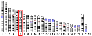
[edit]In human, the SOBP gene is located at the long arm of chromosome 6 at 6q21 and it spans a physical distance of slightly more than 171kbp. The mRNA is transcribed from seven exons, oriented from centromere to telomere, of which the first six exons build the open-reading-frame. The coding mRNA counts 2,622 nucleotides that encode a protein of 873 amino acids.
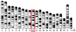
In the mouse, Sopb is located at chromosome 10 at cytogenetic band 10qB2 covering a physical region of 172kbp. As in humans, the mouse Sobp coding region spans six exons but its open-reading-frame is somewhat shorter, counting 2595 nucleotides that encode a protein of 864 amino acids. The protein features two nuclear localization signals on each at its very amino- and carboxy-terminus, two proline-rich sequences in addition to two domains that are related to the FCS-type zinc finger domain. Furthermore, all SOBP proteins share two highly conserved motifs.[7]
Expression
[edit]In the mouse, gene expression profiling by RT-PCR showed a wide expression profile in adult and embryonic tissues with strongest expression being in the brain. By RNA in-situ hybridization, Sobp expression in neonatal tissue was demonstrated in spiral ganglion, the sensory and supporting cells of the maculae of saccule and maculae of utricle, and cristae ampullaris. Sobp is also expressed in the inner nuclear layer of the developing retina at E15, the olfactory epithelium, in neurons of the trigeminal ganglion and in cells surrounding the dermal papillae of hair follicles.
Genetics
[edit]In human, an autosomal recessive mutation causes severe mental retardation with anterior maxillary protrusion and strabismus, named MRAMS syndrome (OMIM #613671). Homozygosity-mapping linked MRAMS syndrome to a 9.8 Mbp region on 6q21. Evaluation of candidate genes within this interval identified a homozygous missense mutation in SOBP in patients with MARMS syndrome. The mutation truncates the SOBP protein near the carboxy-terminus (p.R661X).
In the mouse, two spontaneous recessive autosomal mutations occurred independently at The Jackson Laboratory that were named jackson circler (jc). The first mutation occurred in 1970 on the C57BL/6J background, named C57BL/6J-jc and the second occurred in a B6.129S6 background and was named jc2J. Genetic linkage analyses localized the mutations to chromosome 10. Molecular genetic studies aimed to identify the genetic defect in the jc locus demonstrated a small deletion of 10bp in exon 6 of the Sobp gene. The deletion comprises nucleotides c.1346-1355 and leads to a frame-shift of the open reading frame introducing a stop codon at amino acid position 490 (S449fsX490). In the jc2J allele, the mutation is a nonsense transversion of a guanine to a thymidine (c.1894G>T) changing a glycine to a stop codon (p.G632X).
Phenotypes
[edit]In the mouse, the truncating mutations jc and jc2J lead to profound hearing loss and erratic circling behavior. Specifically, the cochlear duct is shortened, the organ of Corti exhibits supernumerary outer hair cells, mirror image duplications of tunnel of Corti and inner hair cells, as well as ectopic expression of patches of vestibular-like hair cells in Kolliker's organ. The vestibular end organs have a smaller surface area and are thicker.
References
[edit]- ^ a b c GRCh38: Ensembl release 89: ENSG00000112320 – Ensembl, May 2017
- ^ a b c GRCm38: Ensembl release 89: ENSMUSG00000038248 – Ensembl, May 2017
- ^ "Human PubMed Reference:". National Center for Biotechnology Information, U.S. National Library of Medicine.
- ^ "Mouse PubMed Reference:". National Center for Biotechnology Information, U.S. National Library of Medicine.
- ^ "Entrez Gene: sine oculis binding protein homolog (Drosophila)".
- ^ Birk E, Har-Zahav A, Manzini CM, Pasmanik-Chor M, Kornreich L, Walsh CA, et al. (November 2010). "SOBP is mutated in syndromic and nonsyndromic intellectual disability and is highly expressed in the brain limbic system". American Journal of Human Genetics. 87 (5): 694–700. doi:10.1016/j.ajhg.2010.10.005. PMC 2978971. PMID 21035105.
- ^ a b Chen Z, Montcouquiol M, Calderon R, Jenkins NA, Copeland NG, Kelley MW, et al. (June 2008). "Jxc1/Sobp, encoding a nuclear zinc finger protein, is critical for cochlear growth, cell fate, and patterning of the organ of corti". The Journal of Neuroscience. 28 (26): 6633–6641. doi:10.1523/JNEUROSCI.1280-08.2008. PMC 2556235. PMID 18579736.
- ^ Kenyon KL, Li DJ, Clouser C, Tran S, Pignoni F (November 2005). "Fly SIX-type homeodomain proteins Sine oculis and Optix partner with different cofactors during eye development". Dev. Dyn. 234 (3): 497–504. doi:10.1002/dvdy.20442. PMID 15937930. S2CID 11462221.
Further reading
[edit]- Bonaldo MF, Lennon G, Soares MB (September 1996). "Normalization and subtraction: two approaches to facilitate gene discovery". Genome Research. 6 (9): 791–806. doi:10.1101/gr.6.9.791. PMID 8889548.
x* Bonaldo MF, Lennon G, Soares MB (September 1996). "Normalization and subtraction: two approaches to facilitate gene discovery". Genome Research. 6 (9): 791–806. doi:10.1101/gr.6.9.791. PMID 8889548.
- Rose JE, Behm FM, Drgon T, Johnson C, Uhl GR (2010). "Personalized smoking cessation: interactions between nicotine dose, dependence and quit-success genotype score". Molecular Medicine. 16 (7–8): 247–253. doi:10.2119/molmed.2009.00159. PMC 2896464. PMID 20379614.
- Birk E, Har-Zahav A, Manzini CM, Pasmanik-Chor M, Kornreich L, Walsh CA, et al. (November 2010). "SOBP is mutated in syndromic and nonsyndromic intellectual disability and is highly expressed in the brain limbic system". American Journal of Human Genetics. 87 (5): 694–700. doi:10.1016/j.ajhg.2010.10.005. PMC 2978971. PMID 21035105.
- Calderon A, Derr A, Stagner BB, Johnson KR, Martin G, Noben-Trauth K (November 2006). "Cochlear developmental defect and background-dependent hearing thresholds in the Jackson circler (jc) mutant mouse". Hearing Research. 221 (1–2): 44–58. doi:10.1016/j.heares.2006.07.008. PMID 16962269. S2CID 25996561.


