Myosin binding protein C, cardiac


The myosin-binding protein C, cardiac-type is a protein that in humans is encoded by the MYBPC3 gene.[5] This isoform is expressed exclusively in heart muscle during human and mouse development,[6] and is distinct from those expressed in slow skeletal muscle (MYBPC1) and fast skeletal muscle (MYBPC2).
Structure
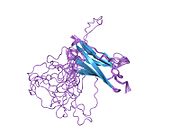
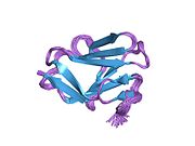
[edit]cMyBP-C is a 140.5 kDa protein composed of 1273 amino acids.[7][8][9] cMyBP-C is a myosin-associated protein that binds at 43 nm intervals along the myosin thick filament backbone, stretching for 200 nm on either side of the M-line within the crossbridge-bearing zone (C-region) of the A band in striated muscle.[10] The approximate stoichiometry of cMyBP-C along the thick filament is 1 per 9-10 myosin molecules, or 37 cMyBP-C molecules per thick filament.[11] In addition to myosin, cMyBP-C also binds titin and actin.[12][13] The cMyBP-C isoform expressed in cardiac muscle differs from those expressed in slow and fast skeletal muscle (MYBPC1 and MYBPC2, respectively) by three features: (1) an additional immunoglobulin (Ig)-like domain on the N-terminus, (2) a linker region between the second and third Ig domains, and (3) an additional loop in the sixth Ig domain.[14] cMyBP-C appears necessary for normal order, filament length and lattice spacing within the structure of the sarcomere.[15][16]
Function
[edit]cMyBP-C is not essential for sarcomere formation during embryogenesis, but is crucial for sarcomere organization and maintenance of normal cardiac function. Absence of cMyBP-C (Mybpc3-targeted knock-out mice) results in severe cardiac hypertrophy, increased heart-weight-to-body-weight-ratios, enlargement of ventricles, increased myofilament Ca2+ sensitivity and depressed diastolic and systolic function.[17][18][19] Histologically, Mybpc3-targeted knock-out hearts display structural rearrangements with cardiac myocyte disarray and increased interstitial fibrosis similar to patients with hypertrophic cardiomyopathy, without obvious alterations in shape or size of single cardiac myocytes. Ultrastructural examination revealed a loss of lateral alignment of adjacent myofibrils with their Z-lines misaligned.[17][18][20][21]
cMyBP-C appears to act as a brake on cardiac contraction, as loaded shortening, power and cycling kinetics all increase in cMyBP-C knockout mice.[22] Consistent with this notion, cMyBP-C knockout mice exhibit an abnormal systolic timecourse, with a shortened elastance timecourse and lower peak elastance in vivo,[23] and an accelerated force development in isolated, skinned cardiac fibers[24] suggesting that cMyBP-C is required to constrain the crossbridges in order to sustain a normal ejection.

cMyBP-C regulates the positioning of myosin and actin for interaction and acts as a tether to the myosin S1 heads, limiting their mobility. This results in a decreased number of crossbridges formed, which hinders force generation, due to its N-terminal C1-M-C2 region interacting with the myosin-S2 domain.[25][26][27][28] Furthermore, cMyBP-C contributes to the regulation of cardiac contraction at short sarcomere length and is required for complete relaxation in diastole.[19][29]
Interactions of cMyBP-C with its binding partners vary with its posttranslational modification status. At least three extensively characterized phosphorylation sites (Ser273, 282 and 302; numbering refers to the mouse sequence) are localized in the M motif of cMyBP-C and are targeted by protein kinases in a hierarchical order of events. In its dephosphorylated state, cMyBP-C binds predominantly to myosin S2 and brakes crossbridge formation, however, when phosphorylated in response to β-adrenergic stimulation through activating cAMP-dependent protein kinase (PKA), it favours binding to actin, then accelerating crossbridge formation, enhancing force development and promoting relaxation.[30] Protein kinases identified thus far to phosphorylate cMyBP-C in the M motif are PKA,[31][32][33][34][35] Ca2+/calmodulin-dependent kinase II (CaMKII),[36] ribosomal s6 kinase (RSK),[37]protein kinase D (PKD),[38][39] and protein kinase C (PKC).[34] Furthermore, GSK3β was described as another protein kinase to phosphorylate cMyBP-C outside the M-domain in the proline-alanine-rich actin-binding site at Ser133 in human myocardium (mouse Ser131).[40] Phosphorylation is required for normal cardiac function and cMyBP-C stability,[41][42] and overall phosphorylation levels of cMyBP-C are reduced in human and experimental heart failure.[43][44] Other posttranslational modifications of cMyBP-C exist, which occur throughout the protein and are not thoroughly characterised yet, such as acetylation,[45] citrullination,[46] S-glutathiolation,[47][48][49][50] S-nitrosylation[51] and carbonylation.[52]
Genetics
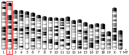
[edit]The cloning of the human MYBPC3 cDNA and localization of the gene on human chromosome 11p11.2 has assisted the structure and function of cMyBP-C.[53] MYBPC3 became therefore the “best” candidate gene for the CMH4 locus for hypertrophic cardiomyopathy that was initially mapped by the group of Schwartz.[54] MYBPC3 mutations segregating in families with hypertrophic cardiomyopathy have been identified.[55][56] MYBPC3 was thus the fourth gene for hypertrophic cardiomyopathy, following MYH7, encoding β-myosin heavy chain, TNNT2 and TPM1, encoding cardiac troponin T and α-tropomyosin, respectively, earmarking hypertrophic cardiomyopathy (HCM) as a disease of the sarcomere. Truncation mutations in MYBPC3 stand as the primary cause of HCM.[57]
To date, roughly 350 mutations in MYBPC3 have been identified, and in large part, the mutations result in protein truncation, shifts in reading frames, and premature termination codons.[58][59] Genetic studies have revealed significant overlap between genotypes and phenotypes as MYBPC3 mutations can lead to various forms of cardiomyopathies, such as dilated cardiomyopathy[60] and left ventricular noncompaction cardiomyopathy.[61] In patients with isolated or familial cases of dilated cardiomyoathy, MYBPC3 mutations represented the second highest number of known mutations.[60] Furthermore, a 25-bp intronic MYBPC3 deletion leading to protein truncation is present in 4% of the population in South India and is associated with a higher risk to develop heart failure.[62] Founder MYBPC3 mutations have been reported in Iceland, Italy, The Netherlands, Japan, France and Finland, where they represent a large percentage of cases with hypertrophic cardiomyopathy. All of them are truncating mutations, resulting in a shorter protein, lacking the regulatory phosphorylatable M motif and/or major binding domains to other sarcomeric proteins.[63][64][65][66][67][68][69] A body of evidence indicates that patients with more than 1 mutation often develop a more severe phenotype,[70] and a significant fraction of childhood-onset hypertrophic cardiomyopathy (14%) is caused by compound genetic variants.[71] This suggests that a gene-dosage effect might be responsible for manifestations at a younger age. A total of 51 cases of homozygotes or compound heterozygotes have been reported, most of them with double truncating MYBPC3 mutations and associated with severe cardiomyopathy, leading to heart failure and death within the first year of life.[72]
Pathomechanisms
[edit]A great understanding of how MYBPC3 mutations lead to the development of inherited cardiomyopathy came from the analyses of human myocardial samples, gene transfer in different cell lines, naturally-occurring or transgenic animal models and more recently disease modeling using induced pluripotent stem cells (iPSC)-derived cardiac myocytes.[73][74] Although access to human myocardial samples is difficult, at least some studies provided evidence that truncated cMyBP-Cs, resulting from truncating MYBPC3 mutations are not detectable in human patient samples by Western-immunoblot analysis.[75][76][77][78] This was supported in heterozygous Mybpc3-targeted knock-in mice,[79] carrying the human c.772G>A transition (i.e. founder mutation in Tuscany[67] These data suggest haploinsufficiency as the main disease mechanism for heterozygous truncating mutations.[80][81] A body of evidence exists that the mechanisms regulating the expression of mutant allele involve the nonsense-mediated mRNA decay, the ubiquitin-proteasome system (UPS) and the autophagy-lysosomal pathway after gene transfer of mutant MYBPC3 in cardiac myocytes or in mice in vivo.[82][83][79][84][85][86] In contrast to truncating mutations, missense mutations lead, in most of the cases (although difficult to specifically detect), to stable mutant cMyBP-Cs that are, at least in part, incorporated into the sarcomere and could act as poison polypeptides on the structure and/or function of the sarcomere. Homozygous or compound heterozygous mutations are therefore likely subject to differential regulation depending on whether they are double missense, double truncating or mixed missense/truncating mutations. The homozygous Mybpc3-targeted knock-in mice, which genetically mimic the situation of severe neonatal cardiomyopathy are born without phenotype and soon after birth develop systolic dysfunction followed by (compensatory) cardiac hypertrophy.[87][88] The human c.772G>A transition results in low levels of three different mutant Mybpc3 mRNAs and cMyBP-Cs in homozygous mice, suggesting a combination of haploinsufficiency and polypeptide poisoning as disease mechanism in the homozygous state.[79] In addition, the combination of external stress (such as neurohumoral stress or aging) and Mybpc3 mutations have been shown to impair the UPS in mice,[89][90] and proteasomal activities were also depressed in patients with hypertrophic cardiomyopathy or dilated cardiomyopathy.[91]
Skinned trabeculae or cardiac myocytes obtained from human patients carrying a MYBPC3 mutation or from heterozygous and homozygous Mybpc3-targeted knock-in mice exhibited higher myofilament Ca2+ sensitivity than controls.[92][78][93][94][95] Disease-modeling by engineered heart tissue (EHT) technology with cardiac cells from heterozygous or homozygous Mybpc3-targeted knock-in mice reproduced observations made in human and mouse studies displaying abbreviated contractions, greater sensitivity to external Ca2+ and smaller inotropic responses to various drugs (isoprenaline, EMD 57033 and verapamil) compared to wild-type control EHTs.[96] Therefore, EHTs are suitable to model the disease phenotype and recapitulate functional alterations found in mice with hypertrophic cardiomyopathy. Another good system for modeling cardiomyopathies in the cell culture dish is the derivation of cardiac myocytes from iPSC. Reports of human iPSC models of sarcomeric cardiomyopathies showed cellular hypertrophy in most of the cases,[97][98][99][100] including one with the c.2995_3010del MYBPC3 mutation that exhibited in addition to hypertrophy contractile variability in the presence of endothelin-1.[100]
Therapy
[edit]Because of their tissue selectivity and persistent expression recombinant adeno-associated viruses (AAV) have therapeutic potential in the treatment of inherited cardiomyopathy resulting from MYBPC3 mutations-[101] Several targeting approaches have been developed.[102][103] The most recent is genome editing to correct a mutation by CRISPR/Cas9 technology.[104] Naturally existing as part of the prokaryotic immune system, the CRISPR/Cas9 system has been used for correction of mutations in the mammalian genome.[105] By inducing nicks in the double-stranded DNA and providing a template DNA sequence, it is possible to repair mutations by homologous recombination. This approach has not yet been evaluated for MYBPC3 mutations, but it could be used for each single or clustered mutation, and therefore applied preferentially for frequent founder MYBPC3 mutations.
Other strategies targeting the mutant pre-mRNA by exon skipping and/or spliceosome-mediated RNA trans-splicing (SMaRT) have been evaluated for MYBPC3. Exon skipping can be achieved using antisense oligonucleotide (AON) masking exonic splicing enhancer sequences and therefore preventing binding of the splicing machinery and therefore resulting in exclusion of the exon from the mRNA.[106][107] This approach can be applied when the resulting shorter, but in-frame translated protein maintains its function. Proof-of-concept of exon skipping was recently shown in homozygous Mybpc3-targeted knock-in mice.[87] Systemic administration of AAV-based AONs to Mybpc3-targeted knock-in newborn mice prevented both systolic dysfunction and left ventricular hypertrophy, at least for the duration of the investigated period.[87] For the human MYBPC3 gene, skipping of 6 single exons or 5 double exons with specific AONs would result in shortened in-frame cMyBP-Cs, allowing the preservation of the functionally important phosphorylation and protein interaction sites. With this approach, about half of missense or exonic/intronic truncating mutations could be removed, including 35 mutations in exon 25. The other strategy targeting the mutant pre-mRNA is SMaRT. Hereby, two independently transcribed molecules, the mutant pre-mRNA and the therapeutic pre-trans-splicing molecule carrying the wild-type sequence are spliced together to give rise to a repaired full-length mRNA.[108] Recently, the feasibility of this method was shown both in isolated cardiac myocytes and in vivo in the heart of homozygous Mybpc3-targeted knock-in mice, although the efficiency of the process was low and the amount of repaired protein was not sufficient to prevent the development of the cardiac disease phenotype.[88] In principle, however, this SmART strategy is superior to exon skipping or CRISPR/Cas9 genome editing and still attractive, because only two pre-trans-splicing molecules, targeting the 5’ and the 3’ of MYBPC3 pre-mRNA would be sufficient to bypass all MYBPC3 mutations associated with cardiomyopathies and therefore repair the mRNA.
AAV-mediated gene transfer of the full-length Mybpc3 (defined as “gene replacement”) dose-dependently prevents the development of cardiac hypertrophy and dysfunction in homozygous Mybpc3-targeted knock-in mice.[109] The dose-dependent expression of exogenous Mybpc3 was associated with the down-regulation of endogenous mutant Mybpc3. Additional expression of a sarcomeric protein is expected to replace partially or completely the endogenous protein level in the sarcomere, as it has been shown in transgenic mice expressing sarcomeric proteins.[73]
Notes
[edit]
The 2015 version of this article was updated by an external expert under a dual publication model. The corresponding academic peer reviewed article was published in Gene and can be cited as: Lucie Carrier, Giulia Mearini, Konstantina Stathopoulou, Friederike Cuello (7 September 2015). "Cardiac myosin-binding protein C (MYBPC3) in cardiac pathophysiology". Gene. Gene Wiki Review Series. 573 (2): 188–197. doi:10.1016/J.GENE.2015.09.008. ISSN 0378-1119. PMC 6660134. PMID 26358504. Wikidata Q38584470. |
References
[edit]- ^ a b c GRCh38: Ensembl release 89: ENSG00000134571 – Ensembl, May 2017
- ^ a b c GRCm38: Ensembl release 89: ENSMUSG00000002100 – Ensembl, May 2017
- ^ "Human PubMed Reference:". National Center for Biotechnology Information, U.S. National Library of Medicine.
- ^ "Mouse PubMed Reference:". National Center for Biotechnology Information, U.S. National Library of Medicine.
- ^ Gautel M, Zuffardi O, Freiburg A, Labeit S (May 1995). "Phosphorylation switches specific for the cardiac isoform of myosin binding protein-C: a modulator of cardiac contraction?". The EMBO Journal. 14 (9): 1952–60. doi:10.1002/j.1460-2075.1995.tb07187.x. PMC 398294. PMID 7744002.
- ^ Fougerousse F, Delezoide AL, Fiszman MY, Schwartz K, Beckmann JS, Carrier L (1998). "Cardiac myosin binding protein C gene is specifically expressed in heart during murine and human development". Circulation Research. 82 (1): 130–3. doi:10.1161/01.res.82.1.130. PMID 9440712.
- ^ Carrier L, Bonne G, Bährend E, Yu B, Richard P, Niel F, Hainque B, Cruaud C, Gary F, Labeit S, Bouhour JB, Dubourg O, Desnos M, Hagège AA, Trent RJ, Komajda M, Fiszman M, Schwartz K (Mar 1997). "Organization and sequence of human cardiac myosin binding protein C gene (MYBPC3) and identification of mutations predicted to produce truncated proteins in familial hypertrophic cardiomyopathy". Circulation Research. 80 (3): 427–34. doi:10.1161/01.res.0000435859.24609.b3. PMID 9048664.
- ^ "Protein Information - Myosin-binding protein C, cardiac-type". Cardiac Organellar Protein Atlas Knowledgebase (COPaKB). NHLBI Proteomics Center at UCLA. Retrieved 29 April 2015.
- ^ Zong NC, Li H, Li H, Lam MP, Jimenez RC, Kim CS, Deng N, Kim AK, Choi JH, Zelaya I, Liem D, Meyer D, Odeberg J, Fang C, Lu HJ, Xu T, Weiss J, Duan H, Uhlen M, Yates JR, Apweiler R, Ge J, Hermjakob H, Ping P (Oct 2013). "Integration of cardiac proteome biology and medicine by a specialized knowledgebase". Circulation Research. 113 (9): 1043–53. doi:10.1161/CIRCRESAHA.113.301151. PMC 4076475. PMID 23965338.
- ^ Bennett P, Craig R, Starr R, Offer G (Dec 1986). "The ultrastructural location of C-protein, X-protein and H-protein in rabbit muscle". Journal of Muscle Research and Cell Motility. 7 (6): 550–67. doi:10.1007/bf01753571. PMID 3543050. S2CID 855781.
- ^ Offer G, Moos C, Starr R (Mar 1973). "A new protein of the thick filaments of vertebrate skeletal myofibrils. Extractions, purification and characterization". Journal of Molecular Biology. 74 (4): 653–76. doi:10.1016/0022-2836(73)90055-7. PMID 4269687.
- ^ Freiburg A, Gautel M (Jan 1996). "A molecular map of the interactions between titin and myosin-binding protein C. Implications for sarcomeric assembly in familial hypertrophic cardiomyopathy". European Journal of Biochemistry. 235 (1–2): 317–23. doi:10.1111/j.1432-1033.1996.00317.x. PMID 8631348.
- ^ Shaffer JF, Kensler RW, Harris SP (May 2009). "The myosin-binding protein C motif binds to F-actin in a phosphorylation-sensitive manner". The Journal of Biological Chemistry. 284 (18): 12318–27. doi:10.1074/jbc.M808850200. PMC 2673300. PMID 19269976.
- ^ Winegrad S (May 1999). "Cardiac myosin binding protein C". Circulation Research. 84 (10): 1117–26. doi:10.1161/01.res.84.10.1117. PMID 10347086.
- ^ Koretz JF (Sep 1979). "Effects of C-protein on synthetic myosin filament structure". Biophysical Journal. 27 (3): 433–46. Bibcode:1979BpJ....27..433K. doi:10.1016/S0006-3495(79)85227-3. PMC 1328598. PMID 263692.
- ^ Colson BA, Bekyarova T, Fitzsimons DP, Irving TC, Moss RL (Mar 2007). "Radial displacement of myosin cross-bridges in mouse myocardium due to ablation of myosin binding protein-C". Journal of Molecular Biology. 367 (1): 36–41. doi:10.1016/j.jmb.2006.12.063. PMC 1892277. PMID 17254601.
- ^ a b Harris SP, Bartley CR, Hacker TA, McDonald KS, Douglas PS, Greaser ML, Powers PA, Moss RL (Mar 2002). "Hypertrophic cardiomyopathy in cardiac myosin binding protein-C knockout mice". Circulation Research. 90 (5): 594–601. doi:10.1161/01.res.0000012222.70819.64. PMID 11909824.
- ^ a b Carrier L, Knöll R, Vignier N, Keller DI, Bausero P, Prudhon B, Isnard R, Ambroisine ML, Fiszman M, Ross J, Schwartz K, Chien KR (Aug 2004). "Asymmetric septal hypertrophy in heterozygous cMyBP-C null mice". Cardiovascular Research. 63 (2): 293–304. doi:10.1016/j.cardiores.2004.04.009. PMID 15249187.
- ^ a b Cazorla O, Szilagyi S, Vignier N, Salazar G, Krämer E, Vassort G, Carrier L, Lacampagne A (Feb 2006). "Length and protein kinase A modulations of myocytes in cardiac myosin binding protein C-deficient mice". Cardiovascular Research. 69 (2): 370–80. doi:10.1016/j.cardiores.2005.11.009. PMID 16380103.
- ^ Brickson S, Fitzsimons DP, Pereira L, Hacker T, Valdivia H, Moss RL (Apr 2007). "In vivo left ventricular functional capacity is compromised in cMyBP-C null mice". American Journal of Physiology. Heart and Circulatory Physiology. 292 (4): H1747–54. doi:10.1152/ajpheart.01037.2006. PMID 17122190.
- ^ Luther PK, Bennett PM, Knupp C, Craig R, Padrón R, Harris SP, Patel J, Moss RL (Dec 2008). "Understanding the organisation and role of myosin binding protein C in normal striated muscle by comparison with MyBP-C knockout cardiac muscle". Journal of Molecular Biology. 384 (1): 60–72. doi:10.1016/j.jmb.2008.09.013. PMC 2593797. PMID 18817784.
- ^ Korte FS, McDonald KS, Harris SP, Moss RL (Oct 2003). "Loaded shortening, power output, and rate of force redevelopment are increased with knockout of cardiac myosin binding protein-C". Circulation Research. 93 (8): 752–8. doi:10.1161/01.RES.0000096363.85588.9A. PMID 14500336.
- ^ Palmer BM, Georgakopoulos D, Janssen PM, Wang Y, Alpert NR, Belardi DF, Harris SP, Moss RL, Burgon PG, Seidman CE, Seidman JG, Maughan DW, Kass DA (May 2004). "Role of cardiac myosin binding protein C in sustaining left ventricular systolic stiffening". Circulation Research. 94 (9): 1249–55. doi:10.1161/01.RES.0000126898.95550.31. PMID 15059932.
- ^ Stelzer JE, Fitzsimons DP, Moss RL (Jun 2006). "Ablation of myosin-binding protein-C accelerates force development in mouse myocardium". Biophysical Journal. 90 (11): 4119–27. Bibcode:2006BpJ....90.4119S. doi:10.1529/biophysj.105.078147. PMC 1459529. PMID 16513777.
- ^ Gruen M, Gautel M (Feb 1999). "Mutations in beta-myosin S2 that cause familial hypertrophic cardiomyopathy (FHC) abolish the interaction with the regulatory domain of myosin-binding protein-C". Journal of Molecular Biology. 286 (3): 933–49. doi:10.1006/jmbi.1998.2522. PMID 10024460.
- ^ Kunst G, Kress KR, Gruen M, Uttenweiler D, Gautel M, Fink RH (2000). "Myosin binding protein C, a phosphorylation-dependent force regulator in muscle that controls the attachment of myosin heads by its interaction with myosin S2". Circulation Research. 86 (1): 51–8. doi:10.1161/01.res.86.1.51. PMID 10625305.
- ^ Harris SP, Rostkova E, Gautel M, Moss RL (Oct 2004). "Binding of myosin binding protein-C to myosin subfragment S2 affects contractility independent of a tether mechanism". Circulation Research. 95 (9): 930–6. doi:10.1161/01.RES.0000147312.02673.56. PMID 15472117.
- ^ Ababou A, Gautel M, Pfuhl M (Mar 2007). "Dissecting the N-terminal myosin binding site of human cardiac myosin-binding protein C. Structure and myosin binding of domain C2". The Journal of Biological Chemistry. 282 (12): 9204–15. doi:10.1074/jbc.M610899200. PMID 17192269.
- ^ Pohlmann L, Kröger I, Vignier N, Schlossarek S, Krämer E, Coirault C, Sultan KR, El-Armouche A, Winegrad S, Eschenhagen T, Carrier L (Oct 2007). "Cardiac myosin-binding protein C is required for complete relaxation in intact myocytes". Circulation Research. 101 (9): 928–38. doi:10.1161/CIRCRESAHA.107.158774. PMID 17823372.
- ^ Moss RL, Fitzsimons DP, Ralphe JC (Jan 2015). "Cardiac MyBP-C regulates the rate and force of contraction in mammalian myocardium". Circulation Research. 116 (1): 183–92. doi:10.1161/CIRCRESAHA.116.300561. PMC 4283578. PMID 25552695.
- ^ Hartzell HC, Titus L (Feb 1982). "Effects of cholinergic and adrenergic agonists on phosphorylation of a 165,000-dalton myofibrillar protein in intact cardiac muscle". The Journal of Biological Chemistry. 257 (4): 2111–20. doi:10.1016/S0021-9258(19)68153-6. PMID 6276407.
- ^ Hartzell HC, Glass DB (Dec 1984). "Phosphorylation of purified cardiac muscle C-protein by purified cAMP-dependent and endogenous Ca2+-calmodulin-dependent protein kinases". The Journal of Biological Chemistry. 259 (24): 15587–96. doi:10.1016/S0021-9258(17)42588-9. PMID 6549009.
- ^ Gautel M, Zuffardi O, Freiburg A, Labeit S (May 1995). "Phosphorylation switches specific for the cardiac isoform of myosin binding protein-C: a modulator of cardiac contraction?". The EMBO Journal. 14 (9): 1952–60. doi:10.1002/j.1460-2075.1995.tb07187.x. PMC 398294. PMID 7744002.
- ^ a b Mohamed AS, Dignam JD, Schlender KK (Oct 1998). "Cardiac myosin-binding protein C (MyBP-C): identification of protein kinase A and protein kinase C phosphorylation sites". Archives of Biochemistry and Biophysics. 358 (2): 313–9. doi:10.1006/abbi.1998.0857. PMID 9784245.
- ^ McClellan G, Kulikovskaya I, Winegrad S (Aug 2001). "Changes in cardiac contractility related to calcium-mediated changes in phosphorylation of myosin-binding protein C". Biophysical Journal. 81 (2): 1083–92. Bibcode:2001BpJ....81.1083M. doi:10.1016/S0006-3495(01)75765-7. PMC 1301577. PMID 11463649.
- ^ Sadayappan S, Gulick J, Osinska H, Barefield D, Cuello F, Avkiran M, Lasko VM, Lorenz JN, Maillet M, Martin JL, Brown JH, Bers DM, Molkentin JD, James J, Robbins J (Jul 2011). "A critical function for Ser-282 in cardiac Myosin binding protein-C phosphorylation and cardiac function". Circulation Research. 109 (2): 141–50. doi:10.1161/CIRCRESAHA.111.242560. PMC 3132348. PMID 21597010.
- ^ Cuello F, Bardswell SC, Haworth RS, Ehler E, Sadayappan S, Kentish JC, Avkiran M (Feb 2011). "Novel role for p90 ribosomal S6 kinase in the regulation of cardiac myofilament phosphorylation". The Journal of Biological Chemistry. 286 (7): 5300–10. doi:10.1074/jbc.M110.202713. PMC 3037642. PMID 21148481.
- ^ Bardswell SC, Cuello F, Rowland AJ, Sadayappan S, Robbins J, Gautel M, Walker JW, Kentish JC, Avkiran M (Feb 2010). "Distinct sarcomeric substrates are responsible for protein kinase D-mediated regulation of cardiac myofilament Ca2+ sensitivity and cross-bridge cycling". The Journal of Biological Chemistry. 285 (8): 5674–82. doi:10.1074/jbc.M109.066456. PMC 2820795. PMID 20018870.
- ^ Dirkx E, Cazorla O, Schwenk RW, Lorenzen-Schmidt I, Sadayappan S, Van Lint J, Carrier L, van Eys GJ, Glatz JF, Luiken JJ (Aug 2012). "Protein kinase D increases maximal Ca2+-activated tension of cardiomyocyte contraction by phosphorylation of cMyBP-C-Ser315". American Journal of Physiology. Heart and Circulatory Physiology. 303 (3): H323–31. doi:10.1152/ajpheart.00749.2011. PMC 6734090. PMID 22636676.
- ^ Kuster DW, Sequeira V, Najafi A, Boontje NM, Wijnker PJ, Witjas-Paalberends ER, Marston SB, Dos Remedios CG, Carrier L, Demmers JA, Redwood C, Sadayappan S, van der Velden J (Feb 2013). "GSK3β phosphorylates newly identified site in the proline-alanine-rich region of cardiac myosin-binding protein C and alters cross-bridge cycling kinetics in human: short communication". Circulation Research. 112 (4): 633–9. doi:10.1161/CIRCRESAHA.112.275602. PMC 3595322. PMID 23277198.
- ^ Govindan S, Sarkey J, Ji X, Sundaresan NR, Gupta MP, de Tombe PP, Sadayappan S (May 2012). "Pathogenic properties of the N-terminal region of cardiac myosin binding protein-C in vitro". Journal of Muscle Research and Cell Motility. 33 (1): 17–30. doi:10.1007/s10974-012-9292-y. PMC 3368277. PMID 22527638.
- ^ Witayavanitkul N, Ait Mou Y, Kuster DW, Khairallah RJ, Sarkey J, Govindan S, Chen X, Ge Y, Rajan S, Wieczorek DF, Irving T, Westfall MV, de Tombe PP, Sadayappan S (Mar 2014). "Myocardial infarction-induced N-terminal fragment of cardiac myosin-binding protein C (cMyBP-C) impairs myofilament function in human myocardium". The Journal of Biological Chemistry. 289 (13): 8818–27. doi:10.1074/jbc.M113.541128. PMC 3979389. PMID 24509847.
- ^ El-Armouche A, Pohlmann L, Schlossarek S, Starbatty J, Yeh YH, Nattel S, Dobrev D, Eschenhagen T, Carrier L (Aug 2007). "Decreased phosphorylation levels of cardiac myosin-binding protein-C in human and experimental heart failure". Journal of Molecular and Cellular Cardiology. 43 (2): 223–9. doi:10.1016/j.yjmcc.2007.05.003. PMID 17560599.
- ^ Copeland O, Sadayappan S, Messer AE, Steinen GJ, van der Velden J, Marston SB (Dec 2010). "Analysis of cardiac myosin binding protein-C phosphorylation in human heart muscle". Journal of Molecular and Cellular Cardiology. 49 (6): 1003–11. doi:10.1016/j.yjmcc.2010.09.007. PMID 20850451.
- ^ Ge Y, Rybakova IN, Xu Q, Moss RL (Aug 2009). "Top-down high-resolution mass spectrometry of cardiac myosin binding protein C revealed that truncation alters protein phosphorylation state". Proceedings of the National Academy of Sciences of the United States of America. 106 (31): 12658–63. Bibcode:2009PNAS..10612658G. doi:10.1073/pnas.0813369106. PMC 2722289. PMID 19541641.
- ^ Fert-Bober J, Sokolove J (Aug 2014). "Proteomics of citrullination in cardiovascular disease". Proteomics: Clinical Applications. 8 (7–8): 522–33. doi:10.1002/prca.201400013. PMID 24946285. S2CID 7008319.
- ^ Brennan JP, Miller JI, Fuller W, Wait R, Begum S, Dunn MJ, Eaton P (Feb 2006). "The utility of N,N-biotinyl glutathione disulfide in the study of protein S-glutathiolation". Molecular & Cellular Proteomics. 5 (2): 215–25. doi:10.1074/mcp.M500212-MCP200. PMID 16223748.
- ^ Lovelock JD, Monasky MM, Jeong EM, Lardin HA, Liu H, Patel BG, Taglieri DM, Gu L, Kumar P, Pokhrel N, Zeng D, Belardinelli L, Sorescu D, Solaro RJ, Dudley SC (Mar 2012). "Ranolazine improves cardiac diastolic dysfunction through modulation of myofilament calcium sensitivity". Circulation Research. 110 (6): 841–50. doi:10.1161/CIRCRESAHA.111.258251. PMC 3314887. PMID 22343711.
- ^ Jeong EM, Monasky MM, Gu L, Taglieri DM, Patel BG, Liu H, Wang Q, Greener I, Dudley SC, Solaro RJ (Mar 2013). "Tetrahydrobiopterin improves diastolic dysfunction by reversing changes in myofilament properties". Journal of Molecular and Cellular Cardiology. 56: 44–54. doi:10.1016/j.yjmcc.2012.12.003. PMC 3666585. PMID 23247392.
- ^ Patel BG, Wilder T, Solaro RJ (2013). "Novel control of cardiac myofilament response to calcium by S-glutathionylation at specific sites of myosin binding protein C". Frontiers in Physiology. 4: 336. doi:10.3389/fphys.2013.00336. PMC 3834529. PMID 24312057.
- ^ Kohr MJ, Aponte AM, Sun J, Wang G, Murphy E, Gucek M, Steenbergen C (Apr 2011). "Characterization of potential S-nitrosylation sites in the myocardium". American Journal of Physiology. Heart and Circulatory Physiology. 300 (4): H1327–35. doi:10.1152/ajpheart.00997.2010. PMC 3075037. PMID 21278135.
- ^ Aryal B, Jeong J, Rao VA (Feb 2014). "Doxorubicin-induced carbonylation and degradation of cardiac myosin binding protein C promote cardiotoxicity". Proceedings of the National Academy of Sciences of the United States of America. 111 (5): 2011–6. Bibcode:2014PNAS..111.2011A. doi:10.1073/pnas.1321783111. PMC 3918758. PMID 24449919.
- ^ Gautel M, Zuffardi O, Freiburg A, Labeit S (May 1995). "Phosphorylation switches specific for the cardiac isoform of myosin binding protein-C: a modulator of cardiac contraction?". The EMBO Journal. 14 (9): 1952–60. doi:10.1002/j.1460-2075.1995.tb07187.x. PMC 398294. PMID 7744002.
- ^ Carrier L, Hengstenberg C, Beckmann JS, Guicheney P, Dufour C, Bercovici J, Dausse E, Berebbi-Bertrand I, Wisnewsky C, Pulvenis D (Jul 1993). "Mapping of a novel gene for familial hypertrophic cardiomyopathy to chromosome 11". Nature Genetics. 4 (3): 311–3. doi:10.1038/ng0793-311. PMID 8358441. S2CID 7535967.
- ^ Bonne G, Carrier L, Bercovici J, Cruaud C, Richard P, Hainque B, Gautel M, Labeit S, James M, Beckmann J, Weissenbach J, Vosberg HP, Fiszman M, Komajda M, Schwartz K (Dec 1995). "Cardiac myosin binding protein-C gene splice acceptor site mutation is associated with familial hypertrophic cardiomyopathy". Nature Genetics. 11 (4): 438–40. doi:10.1038/ng1295-438. PMID 7493026. S2CID 11679535.
- ^ Watkins H, Conner D, Thierfelder L, Jarcho JA, MacRae C, McKenna WJ, Maron BJ, Seidman JG, Seidman CE (Dec 1995). "Mutations in the cardiac myosin binding protein-C gene on chromosome 11 cause familial hypertrophic cardiomyopathy". Nature Genetics. 11 (4): 434–7. doi:10.1038/ng1295-434. PMID 7493025. S2CID 25615613.
- ^ O'Leary TS, Snyder J, Sadayappan S, Day SM, Previs MJ (2019). "MYBPC3 truncation mutations enhance actomyosin contractile mechanics in human hypertrophic cardiomyopathy". Journal of Molecular and Cellular Cardiology. 127: 165–173. doi:10.1016/j.yjmcc.2018.12.003. ISSN 0022-2828. PMC 6592272. PMID 30550750.
- ^ Harris SP, Lyons RG, Bezold KL (Mar 2011). "In the thick of it: HCM-causing mutations in myosin binding proteins of the thick filament". Circulation Research. 108 (6): 751–64. doi:10.1161/CIRCRESAHA.110.231670. PMC 3076008. PMID 21415409.
- ^ Behrens-Gawlik V, Mearini G, Gedicke-Hornung C, Richard P, Carrier L (Feb 2014). "MYBPC3 in hypertrophic cardiomyopathy: from mutation identification to RNA-based correction". Pflügers Archiv. 466 (2): 215–23. doi:10.1007/s00424-013-1409-7. PMID 24337823. S2CID 6625266.
- ^ a b Haas J, Frese KS, Peil B, Kloos W, Keller A, Nietsch R, Feng Z, Müller S, Kayvanpour E, Vogel B, Sedaghat-Hamedani F, Lim WK, Zhao X, Fradkin D, Köhler D, Fischer S, Franke J, Marquart S, Barb I, Li DT, Amr A, Ehlermann P, Mereles D, Weis T, Hassel S, Kremer A, King V, Wirsz E, Isnard R, Komajda M, Serio A, Grasso M, Syrris P, Wicks E, Plagnol V, Lopes L, Gadgaard T, Eiskjær H, Jørgensen M, Garcia-Giustiniani D, Ortiz-Genga M, Crespo-Leiro MG, Deprez RH, Christiaans I, van Rijsingen IA, Wilde AA, Waldenstrom A, Bolognesi M, Bellazzi R, Mörner S, Bermejo JL, Monserrat L, Villard E, Mogensen J, Pinto YM, Charron P, Elliott P, Arbustini E, Katus HA, Meder B (May 2015). "Atlas of the clinical genetics of human dilated cardiomyopathy". European Heart Journal. 36 (18): 1123–35. doi:10.1093/eurheartj/ehu301. hdl:2183/19982. PMID 25163546.
- ^ Probst S, Oechslin E, Schuler P, Greutmann M, Boyé P, Knirsch W, Berger F, Thierfelder L, Jenni R, Klaassen S (Aug 2011). "Sarcomere gene mutations in isolated left ventricular noncompaction cardiomyopathy do not predict clinical phenotype". Circulation: Cardiovascular Genetics. 4 (4): 367–74. doi:10.1161/CIRCGENETICS.110.959270. PMID 21551322.
- ^ Dhandapany PS, Sadayappan S, Xue Y, Powell GT, Rani DS, Nallari P, Rai TS, Khullar M, Soares P, Bahl A, Tharkan JM, Vaideeswar P, Rathinavel A, Narasimhan C, Ayapati DR, Ayub Q, Mehdi SQ, Oppenheimer S, Richards MB, Price AL, Patterson N, Reich D, Singh L, Tyler-Smith C, Thangaraj K (Feb 2009). "A common MYBPC3 (cardiac myosin binding protein C) variant associated with cardiomyopathies in South Asia". Nature Genetics. 41 (2): 187–91. doi:10.1038/ng.309. PMC 2697598. PMID 19151713.
- ^ Adalsteinsdottir B, Teekakirikul P, Maron BJ, Burke MA, Gudbjartsson DF, Holm H, Stefansson K, DePalma SR, Mazaika E, McDonough B, Danielsen R, Seidman JG, Seidman CE, Gunnarsson GT (Sep 2014). "Nationwide study on hypertrophic cardiomyopathy in Iceland: evidence of a MYBPC3 founder mutation". Circulation. 130 (14): 1158–67. doi:10.1161/CIRCULATIONAHA.114.011207. PMID 25078086.
- ^ Calore C, De Bortoli M, Romualdi C, Lorenzon A, Angelini A, Basso C, Thiene G, Iliceto S, Rampazzo A, Melacini P (May 2015). "A founder MYBPC3 mutation results in HCM with a high risk of sudden death after the fourth decade of life". Journal of Medical Genetics. 52 (5): 338–47. doi:10.1136/jmedgenet-2014-102923. PMID 25740977. S2CID 35343228.
- ^ Christiaans I, Nannenberg EA, Dooijes D, Jongbloed RJ, Michels M, Postema PG, Majoor-Krakauer D, van den Wijngaard A, Mannens MM, van Tintelen JP, van Langen IM, Wilde AA (May 2010). "Founder mutations in hypertrophic cardiomyopathy patients in the Netherlands". Netherlands Heart Journal. 18 (5): 248–54. doi:10.1007/bf03091771. PMC 2871745. PMID 20505798.
- ^ Kubo T, Kitaoka H, Okawa M, Matsumura Y, Hitomi N, Yamasaki N, Furuno T, Takata J, Nishinaga M, Kimura A, Doi YL (Nov 2005). "Lifelong left ventricular remodeling of hypertrophic cardiomyopathy caused by a founder frameshift deletion mutation in the cardiac Myosin-binding protein C gene among Japanese". Journal of the American College of Cardiology. 46 (9): 1737–43. doi:10.1016/j.jacc.2005.05.087. PMID 16256878.
- ^ a b Girolami F, Olivotto I, Passerini I, Zachara E, Nistri S, Re F, Fantini S, Baldini K, Torricelli F, Cecchi F (Aug 2006). "A molecular screening strategy based on beta-myosin heavy chain, cardiac myosin binding protein C and troponin T genes in Italian patients with hypertrophic cardiomyopathy". Journal of Cardiovascular Medicine. 7 (8): 601–7. doi:10.2459/01.JCM.0000237908.26377.d6. PMID 16858239. S2CID 20926873.
- ^ Teirlinck CH, Senni F, Malti RE, Majoor-Krakauer D, Fellmann F, Millat G, André-Fouët X, Pernot F, Stumpf M, Boutarin J, Bouvagnet P (2012). "A human MYBPC3 mutation appearing about 10 centuries ago results in a hypertrophic cardiomyopathy with delayed onset, moderate evolution but with a risk of sudden death". BMC Medical Genetics. 13: 105. doi:10.1186/1471-2350-13-105. PMC 3549277. PMID 23140321.
- ^ Jääskeläinen P, Miettinen R, Kärkkäinen P, Toivonen L, Laakso M, Kuusisto J (2004). "Genetics of hypertrophic cardiomyopathy in eastern Finland: few founder mutations with benign or intermediary phenotypes". Annals of Medicine. 36 (1): 23–32. doi:10.1080/07853890310017161. PMID 15000344. S2CID 29985750.
- ^ Richard P, Charron P, Carrier L, Ledeuil C, Cheav T, Pichereau C, Benaiche A, Isnard R, Dubourg O, Burban M, Gueffet JP, Millaire A, Desnos M, Schwartz K, Hainque B, Komajda M (May 2003). "Hypertrophic cardiomyopathy: distribution of disease genes, spectrum of mutations, and implications for a molecular diagnosis strategy". Circulation. 107 (17): 2227–32. doi:10.1161/01.CIR.0000066323.15244.54. PMID 12707239.
- ^ Morita H, Rehm HL, Menesses A, McDonough B, Roberts AE, Kucherlapati R, Towbin JA, Seidman JG, Seidman CE (May 2008). "Shared genetic causes of cardiac hypertrophy in children and adults". The New England Journal of Medicine. 358 (18): 1899–908. doi:10.1056/NEJMoa075463. PMC 2752150. PMID 18403758.
- ^ Wessels MW, Herkert JC, Frohn-Mulder IM, Dalinghaus M, van den Wijngaard A, de Krijger RR, Michels M, de Coo IF, Hoedemaekers YM, Dooijes D (Oct 2014). "Compound heterozygous or homozygous truncating MYBPC3 mutations cause lethal cardiomyopathy with features of noncompaction and septal defects". European Journal of Human Genetics. 23 (7): 922–8. doi:10.1038/ejhg.2014.211. PMC 4463499. PMID 25335496.
- ^ a b Duncker DJ, Bakkers J, Brundel BJ, Robbins J, Tardiff JC, Carrier L (Apr 2015). "Animal and in silico models for the study of sarcomeric cardiomyopathies". Cardiovascular Research. 105 (4): 439–48. doi:10.1093/cvr/cvv006. PMC 4375391. PMID 25600962.
- ^ Eschenhagen T, Mummery C, Knollmann BC (Apr 2015). "Modelling sarcomeric cardiomyopathies in the dish: from human heart samples to iPSC cardiomyocytes". Cardiovascular Research. 105 (4): 424–38. doi:10.1093/cvr/cvv017. PMC 4349163. PMID 25618410.
- ^ Rottbauer W, Gautel M, Zehelein J, Labeit S, Franz WM, Fischer C, Vollrath B, Mall G, Dietz R, Kübler W, Katus HA (Jul 1997). "Novel splice donor site mutation in the cardiac myosin-binding protein-C gene in familial hypertrophic cardiomyopathy. Characterization Of cardiac transcript and protein". The Journal of Clinical Investigation. 100 (2): 475–82. doi:10.1172/JCI119555. PMC 508212. PMID 9218526.
- ^ Moolman JA, Reith S, Uhl K, Bailey S, Gautel M, Jeschke B, Fischer C, Ochs J, McKenna WJ, Klues H, Vosberg HP (Mar 2000). "A newly created splice donor site in exon 25 of the MyBP-C gene is responsible for inherited hypertrophic cardiomyopathy with incomplete disease penetrance". Circulation. 101 (12): 1396–402. doi:10.1161/01.cir.101.12.1396. PMID 10736283.
- ^ Marston S, Copeland O, Jacques A, Livesey K, Tsang V, McKenna WJ, Jalilzadeh S, Carballo S, Redwood C, Watkins H (Jul 2009). "Evidence from human myectomy samples that MYBPC3 mutations cause hypertrophic cardiomyopathy through haploinsufficiency". Circulation Research. 105 (3): 219–22. doi:10.1161/CIRCRESAHA.109.202440. hdl:10044/1/19192. PMID 19574547.
- ^ a b van Dijk SJ, Dooijes D, dos Remedios C, Michels M, Lamers JM, Winegrad S, Schlossarek S, Carrier L, ten Cate FJ, Stienen GJ, van der Velden J (Mar 2009). "Cardiac myosin-binding protein C mutations and hypertrophic cardiomyopathy: haploinsufficiency, deranged phosphorylation, and cardiomyocyte dysfunction". Circulation. 119 (11): 1473–83. doi:10.1161/CIRCULATIONAHA.108.838672. PMID 19273718.
- ^ a b c Vignier N, Schlossarek S, Fraysse B, Mearini G, Krämer E, Pointu H, Mougenot N, Guiard J, Reimer R, Hohenberg H, Schwartz K, Vernet M, Eschenhagen T, Carrier L (Jul 2009). "Nonsense-mediated mRNA decay and ubiquitin-proteasome system regulate cardiac myosin-binding protein C mutant levels in cardiomyopathic mice". Circulation Research. 105 (3): 239–48. doi:10.1161/CIRCRESAHA.109.201251. PMID 19590044.
- ^ Marston S, Copeland O, Gehmlich K, Schlossarek S, Carrier L, Carrier L (May 2012). "How do MYBPC3 mutations cause hypertrophic cardiomyopathy?". Journal of Muscle Research and Cell Motility. 33 (1): 75–80. doi:10.1007/s10974-011-9268-3. PMID 22057632. S2CID 10978237.
- ^ van der Velden J, Ho CY, Tardiff JC, Olivotto I, Knollmann BC, Carrier L (Apr 2015). "Research priorities in sarcomeric cardiomyopathies". Cardiovascular Research. 105 (4): 449–56. doi:10.1093/cvr/cvv019. PMC 4375392. PMID 25631582.
- ^ Sarikas A, Carrier L, Schenke C, Doll D, Flavigny J, Lindenberg KS, Eschenhagen T, Zolk O (Apr 2005). "Impairment of the ubiquitin-proteasome system by truncated cardiac myosin binding protein C mutants". Cardiovascular Research. 66 (1): 33–44. doi:10.1016/j.cardiores.2005.01.004. PMID 15769446.
- ^ Bahrudin U, Morisaki H, Morisaki T, Ninomiya H, Higaki K, Nanba E, Igawa O, Takashima S, Mizuta E, Miake J, Yamamoto Y, Shirayoshi Y, Kitakaze M, Carrier L, Hisatome I (Dec 2008). "Ubiquitin-proteasome system impairment caused by a missense cardiac myosin-binding protein C mutation and associated with cardiac dysfunction in hypertrophic cardiomyopathy" (PDF). Journal of Molecular Biology. 384 (4): 896–907. doi:10.1016/j.jmb.2008.09.070. PMID 18929575.
- ^ Mearini G, Schlossarek S, Willis MS, Carrier L (Dec 2008). "The ubiquitin-proteasome system in cardiac dysfunction" (PDF). Biochimica et Biophysica Acta (BBA) - Molecular Basis of Disease. 1782 (12): 749–63. doi:10.1016/j.bbadis.2008.06.009. PMID 18634872. S2CID 14570410.
- ^ Carrier L, Schlossarek S, Willis MS, Eschenhagen T (Jan 2010). "The ubiquitin-proteasome system and nonsense-mediated mRNA decay in hypertrophic cardiomyopathy". Cardiovascular Research. 85 (2): 330–8. doi:10.1093/cvr/cvp247. PMC 4023315. PMID 19617224.
- ^ Schlossarek S, Frey N, Carrier L (Jun 2014). "Ubiquitin-proteasome system and hereditary cardiomyopathies". Journal of Molecular and Cellular Cardiology. 71: 25–31. doi:10.1016/j.yjmcc.2013.12.016. PMID 24380728.
- ^ a b c Gedicke-Hornung C, Behrens-Gawlik V, Reischmann S, Geertz B, Stimpel D, Weinberger F, Schlossarek S, Précigout G, Braren I, Eschenhagen T, Mearini G, Lorain S, Voit T, Dreyfus PA, Garcia L, Carrier L (Jul 2013). "Rescue of cardiomyopathy through U7snRNA-mediated exon skipping in Mybpc3-targeted knock-in mice". EMBO Molecular Medicine. 5 (7): 1128–1145. doi:10.1002/emmm.201202168. PMC 3721478. PMID 23716398.
- ^ a b Mearini G, Stimpel D, Krämer E, Geertz B, Braren I, Gedicke-Hornung C, Précigout G, Müller OJ, Katus HA, Eschenhagen T, Voit T, Garcia L, Lorain S, Carrier L (2013). "Repair of Mybpc3 mRNA by 5'-trans-splicing in a Mouse Model of Hypertrophic Cardiomyopathy". Molecular Therapy: Nucleic Acids. 2 (7): e102. doi:10.1038/mtna.2013.31. PMC 3731888. PMID 23820890.
- ^ Schlossarek S, Englmann DR, Sultan KR, Sauer M, Eschenhagen T, Carrier L (Jan 2012). "Defective proteolytic systems in Mybpc3-targeted mice with cardiac hypertrophy". Basic Research in Cardiology. 107 (1): 235. doi:10.1007/s00395-011-0235-3. PMID 22189562. S2CID 6472866.
- ^ Schlossarek S, Schuermann F, Geertz B, Mearini G, Eschenhagen T, Carrier L (May 2012). "Adrenergic stress reveals septal hypertrophy and proteasome impairment in heterozygous Mybpc3-targeted knock-in mice". Journal of Muscle Research and Cell Motility. 33 (1): 5–15. doi:10.1007/s10974-011-9273-6. PMID 22076249. S2CID 17638722.
- ^ Predmore JM, Wang P, Davis F, Bartolone S, Westfall MV, Dyke DB, Pagani F, Powell SR, Day SM (Mar 2010). "Ubiquitin proteasome dysfunction in human hypertrophic and dilated cardiomyopathies". Circulation. 121 (8): 997–1004. doi:10.1161/CIRCULATIONAHA.109.904557. PMC 2857348. PMID 20159828.
- ^ Witt CC, Gerull B, Davies MJ, Centner T, Linke WA, Thierfelder L (Feb 2001). "Hypercontractile properties of cardiac muscle fibers in a knock-in mouse model of cardiac myosin-binding protein-C". The Journal of Biological Chemistry. 276 (7): 5353–9. doi:10.1074/jbc.M008691200. PMID 11096095.
- ^ Fraysse B, Weinberger F, Bardswell SC, Cuello F, Vignier N, Geertz B, Starbatty J, Krämer E, Coirault C, Eschenhagen T, Kentish JC, Avkiran M, Carrier L (Jun 2012). "Increased myofilament Ca2+ sensitivity and diastolic dysfunction as early consequences of Mybpc3 mutation in heterozygous knock-in mice". Journal of Molecular and Cellular Cardiology. 52 (6): 1299–307. doi:10.1016/j.yjmcc.2012.03.009. PMC 3370652. PMID 22465693.
- ^ van Dijk SJ, Paalberends ER, Najafi A, Michels M, Sadayappan S, Carrier L, Boontje NM, Kuster DW, van Slegtenhorst M, Dooijes D, dos Remedios C, ten Cate FJ, Stienen GJ, van der Velden J (Jan 2012). "Contractile dysfunction irrespective of the mutant protein in human hypertrophic cardiomyopathy with normal systolic function". Circulation: Heart Failure. 5 (1): 36–46. doi:10.1161/CIRCHEARTFAILURE.111.963702. PMID 22178992.
- ^ Sequeira V, Wijnker PJ, Nijenkamp LL, Kuster DW, Najafi A, Witjas-Paalberends ER, Regan JA, Boontje N, Ten Cate FJ, Germans T, Carrier L, Sadayappan S, van Slegtenhorst MA, Zaremba R, Foster DB, Murphy AM, Poggesi C, Dos Remedios C, Stienen GJ, Ho CY, Michels M, van der Velden J (May 2013). "Perturbed length-dependent activation in human hypertrophic cardiomyopathy with missense sarcomeric gene mutations". Circulation Research. 112 (11): 1491–505. doi:10.1161/CIRCRESAHA.111.300436. PMC 3675884. PMID 23508784.
- ^ Stöhr A, Friedrich FW, Flenner F, Geertz B, Eder A, Schaaf S, Hirt MN, Uebeler J, Schlossarek S, Carrier L, Hansen A, Eschenhagen T (Oct 2013). "Contractile abnormalities and altered drug response in engineered heart tissue from Mybpc3-targeted knock-in mice". Journal of Molecular and Cellular Cardiology. 63: 189–98. doi:10.1016/j.yjmcc.2013.07.011. PMID 23896226.
- ^ Jung G, Bernstein D (Jul 2014). "hiPSC Modeling of Inherited Cardiomyopathies". Current Treatment Options in Cardiovascular Medicine. 16 (7): 320. doi:10.1007/s11936-014-0320-7. PMC 4096486. PMID 24838688.
- ^ Lan F, Lee AS, Liang P, Sanchez-Freire V, Nguyen PK, Wang L, Han L, Yen M, Wang Y, Sun N, Abilez OJ, Hu S, Ebert AD, Navarrete EG, Simmons CS, Wheeler M, Pruitt B, Lewis R, Yamaguchi Y, Ashley EA, Bers DM, Robbins RC, Longaker MT, Wu JC (Jan 2013). "Abnormal calcium handling properties underlie familial hypertrophic cardiomyopathy pathology in patient-specific induced pluripotent stem cells". Cell Stem Cell. 12 (1): 101–13. doi:10.1016/j.stem.2012.10.010. PMC 3638033. PMID 23290139.
- ^ Han L, Li Y, Tchao J, Kaplan AD, Lin B, Li Y, Mich-Basso J, Lis A, Hassan N, London B, Bett GC, Tobita K, Rasmusson RL, Yang L (Nov 2014). "Study familial hypertrophic cardiomyopathy using patient-specific induced pluripotent stem cells". Cardiovascular Research. 104 (2): 258–69. doi:10.1093/cvr/cvu205. PMC 4217687. PMID 25209314.
- ^ a b Tanaka A, Yuasa S, Mearini G, Egashira T, Seki T, Kodaira M, Kusumoto D, Kuroda Y, Okata S, Suzuki T, Inohara T, Arimura T, Makino S, Kimura K, Kimura A, Furukawa T, Carrier L, Node K, Fukuda K (Dec 2014). "Endothelin-1 induces myofibrillar disarray and contractile vector variability in hypertrophic cardiomyopathy-induced pluripotent stem cell-derived cardiomyocytes". Journal of the American Heart Association. 3 (6): e001263. doi:10.1161/JAHA.114.001263. PMC 4338713. PMID 25389285.
- ^ Zacchigna S, Zentilin L, Giacca M (May 2014). "Adeno-associated virus vectors as therapeutic and investigational tools in the cardiovascular system". Circulation Research. 114 (11): 1827–46. doi:10.1161/CIRCRESAHA.114.302331. PMID 24855205.
- ^ Hammond SM, Wood MJ (May 2011). "Genetic therapies for RNA mis-splicing diseases". Trends in Genetics. 27 (5): 196–205. doi:10.1016/j.tig.2011.02.004. PMID 21497936.
- ^ Doudna JA, Charpentier E (Nov 2014). "Genome editing. The new frontier of genome engineering with CRISPR-Cas9". Science. 346 (6213): 1258096. doi:10.1126/science.1258096. PMID 25430774. S2CID 6299381.
- ^ Hsu PD, Lander ES, Zhang F (Jun 2014). "Development and applications of CRISPR-Cas9 for genome engineering". Cell. 157 (6): 1262–78. doi:10.1016/j.cell.2014.05.010. PMC 4343198. PMID 24906146.
- ^ Ran FA, Hsu PD, Wright J, Agarwala V, Scott DA, Zhang F (Nov 2013). "Genome engineering using the CRISPR-Cas9 system". Nature Protocols. 8 (11): 2281–308. doi:10.1038/nprot.2013.143. PMC 3969860. PMID 24157548.
- ^ Woodley L, Valcárcel J (Oct 2002). "Regulation of alternative pre-mRNA splicing". Briefings in Functional Genomics & Proteomics. 1 (3): 266–77. doi:10.1093/bfgp/1.3.266. PMID 15239893.
- ^ Goyenvalle A, Vulin A, Fougerousse F, Leturcq F, Kaplan JC, Garcia L, Danos O (Dec 2004). "Rescue of dystrophic muscle through U7 snRNA-mediated exon skipping". Science. 306 (5702): 1796–9. Bibcode:2004Sci...306.1796G. doi:10.1126/science.1104297. PMID 15528407. S2CID 9359783.
- ^ Wally V, Murauer EM, Bauer JW (Aug 2012). "Spliceosome-mediated trans-splicing: the therapeutic cut and paste". The Journal of Investigative Dermatology. 132 (8): 1959–66. doi:10.1038/jid.2012.101. PMID 22495179.
- ^ Mearini G, Stimpel D, Geertz B, Weinberger F, Krämer E, Schlossarek S, Mourot-Filiatre J, Stoehr A, Dutsch A, Wijnker PJ, Braren I, Katus HA, Müller OJ, Voit T, Eschenhagen T, Carrier L (2014). "Mybpc3 gene therapy for neonatal cardiomyopathy enables long-term disease prevention in mice". Nature Communications. 5: 5515. Bibcode:2014NatCo...5.5515M. doi:10.1038/ncomms6515. PMID 25463264.
Further reading
[edit]- Vikstrom KL, Leinwand LA (Feb 1996). "Contractile protein mutations and heart disease". Current Opinion in Cell Biology. 8 (1): 97–105. doi:10.1016/S0955-0674(96)80053-6. PMID 8791411.
- Schaub MC, Hefti MA, Zuellig RA, Morano I (Feb 1998). "Modulation of contractility in human cardiac hypertrophy by myosin essential light chain isoforms" (PDF). Cardiovascular Research. 37 (2): 381–404. doi:10.1016/S0008-6363(97)00258-7. PMID 9614495.
- Bonne G, Carrier L, Richard P, Hainque B, Schwartz K (Sep 1998). "Familial hypertrophic cardiomyopathy: from mutations to functional defects". Circulation Research. 83 (6): 580–93. doi:10.1161/01.res.83.6.580. PMID 9742053.
- Jääskeläinen P, Miettinen R, Kärkkäinen P, Toivonen L, Laakso M, Kuusisto J (2004). "Genetics of hypertrophic cardiomyopathy in eastern Finland: few founder mutations with benign or intermediary phenotypes". Annals of Medicine. 36 (1): 23–32. doi:10.1080/07853890310017161. PMID 15000344. S2CID 29985750.
- Starr R, Offer G (Jun 1978). "The interaction of C-protein with heavy meromyosin and subfragment-2". The Biochemical Journal. 171 (3): 813–6. doi:10.1042/bj1710813. PMC 1184031. PMID 352343.
- Moos C, Feng IN (Oct 1980). "Effect of C-protein on actomyosin ATPase". Biochimica et Biophysica Acta (BBA) - General Subjects. 632 (2): 141–9. doi:10.1016/0304-4165(80)90071-9. PMID 6448079.
- Watkins H, Conner D, Thierfelder L, Jarcho JA, MacRae C, McKenna WJ, Maron BJ, Seidman JG, Seidman CE (Dec 1995). "Mutations in the cardiac myosin binding protein-C gene on chromosome 11 cause familial hypertrophic cardiomyopathy". Nature Genetics. 11 (4): 434–7. doi:10.1038/ng1295-434. PMID 7493025. S2CID 25615613.
- Bonne G, Carrier L, Bercovici J, Cruaud C, Richard P, Hainque B, Gautel M, Labeit S, James M, Beckmann J, Weissenbach J, Vosberg HP, Fiszman M, Komajda M, Schwartz K (Dec 1995). "Cardiac myosin binding protein-C gene splice acceptor site mutation is associated with familial hypertrophic cardiomyopathy". Nature Genetics. 11 (4): 438–40. doi:10.1038/ng1295-438. PMID 7493026. S2CID 11679535.
- Gautel M, Zuffardi O, Freiburg A, Labeit S (May 1995). "Phosphorylation switches specific for the cardiac isoform of myosin binding protein-C: a modulator of cardiac contraction?". The EMBO Journal. 14 (9): 1952–60. doi:10.1002/j.1460-2075.1995.tb07187.x. PMC 398294. PMID 7744002.
- Carrier L, Hengstenberg C, Beckmann JS, Guicheney P, Dufour C, Bercovici J, Dausse E, Berebbi-Bertrand I, Wisnewsky C, Pulvenis D (Jul 1993). "Mapping of a novel gene for familial hypertrophic cardiomyopathy to chromosome 11". Nature Genetics. 4 (3): 311–3. doi:10.1038/ng0793-311. PMID 8358441. S2CID 7535967.
- Freiburg A, Gautel M (Jan 1996). "A molecular map of the interactions between titin and myosin-binding protein C. Implications for sarcomeric assembly in familial hypertrophic cardiomyopathy". European Journal of Biochemistry. 235 (1–2): 317–23. doi:10.1111/j.1432-1033.1996.00317.x. PMID 8631348.
- Carrier L, Bonne G, Bährend E, Yu B, Richard P, Niel F, Hainque B, Cruaud C, Gary F, Labeit S, Bouhour JB, Dubourg O, Desnos M, Hagège AA, Trent RJ, Komajda M, Fiszman M, Schwartz K (Mar 1997). "Organization and sequence of human cardiac myosin binding protein C gene (MYBPC3) and identification of mutations predicted to produce truncated proteins in familial hypertrophic cardiomyopathy". Circulation Research. 80 (3): 427–34. doi:10.1161/01.res.0000435859.24609.b3. PMID 9048664.
- Rottbauer W, Gautel M, Zehelein J, Labeit S, Franz WM, Fischer C, Vollrath B, Mall G, Dietz R, Kübler W, Katus HA (Jul 1997). "Novel splice donor site mutation in the cardiac myosin-binding protein-C gene in familial hypertrophic cardiomyopathy. Characterization Of cardiac transcript and protein". The Journal of Clinical Investigation. 100 (2): 475–82. doi:10.1172/JCI119555. PMC 508212. PMID 9218526.
- Yu B, French JA, Carrier L, Jeremy RW, McTaggart DR, Nicholson MR, Hambly B, Semsarian C, Richmond DR, Schwartz K, Trent RJ (Mar 1998). "Molecular pathology of familial hypertrophic cardiomyopathy caused by mutations in the cardiac myosin binding protein C gene". Journal of Medical Genetics. 35 (3): 205–10. doi:10.1136/jmg.35.3.205. PMC 1051243. PMID 9541104.
- Moolman-Smook JC, Mayosi B, Brink P, Corfield VA (Mar 1998). "Identification of a new missense mutation in MyBP-C associated with hypertrophic cardiomyopathy". Journal of Medical Genetics. 35 (3): 253–4. doi:10.1136/jmg.35.3.253. PMC 1051254. PMID 9541115.
- Niimura H, Bachinski LL, Sangwatanaroj S, Watkins H, Chudley AE, McKenna W, Kristinsson A, Roberts R, Sole M, Maron BJ, Seidman JG, Seidman CE (Apr 1998). "Mutations in the gene for cardiac myosin-binding protein C and late-onset familial hypertrophic cardiomyopathy". The New England Journal of Medicine. 338 (18): 1248–57. doi:10.1056/NEJM199804303381802. PMID 9562578.
- Richard P, Isnard R, Carrier L, Dubourg O, Donatien Y, Mathieu B, Bonne G, Gary F, Charron P, Hagege M, Komajda M, Schwartz K, Hainque B (Jul 1999). "Double heterozygosity for mutations in the beta-myosin heavy chain and in the cardiac myosin binding protein C genes in a family with hypertrophic cardiomyopathy". Journal of Medical Genetics. 36 (7): 542–5. doi:10.1136/jmg.36.7.542. PMC 1734410. PMID 10424815.
External links
[edit]- Mass spectrometry characterization of MYBPC3 at COPaKB
- GeneReviews/NIH/NCBI/UW entry on Familial Hypertrophic Cardiomyopathy Overview
- Overview of all the structural information available in the PDB for UniProt: Q14896 (Myosin-binding protein C, cardiac-type) at the PDBe-KB.


