Jump to content

File:Wuchereria bancrofti LifeCycle.gif

Page contents not supported in other languages.
This is a file from the Wikimedia Commons
From Wikipedia, the free encyclopedia

Wuchereria_bancrofti_LifeCycle.gif (513 × 435 pixels, file size: 33 KB, MIME type: image/gif)

Description

Wuchereria bancrofti

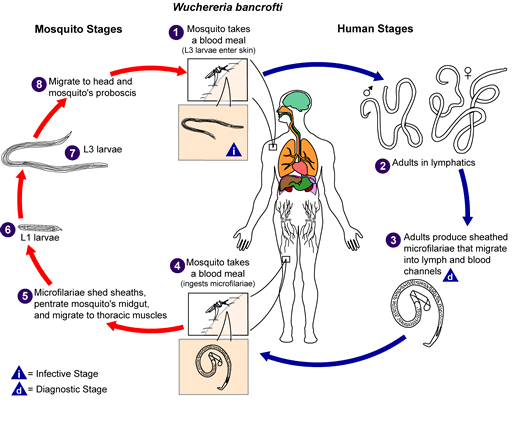
Life cycle of Wuchereria bancrofti

Different species of the following genera of mosquitoes are vectors of W. bancrofti filariasis depending on geographical distribution. Among them are: Culex (C. annulirostris, C. bitaeniorhynchus, C. quinquefasciatus, and C. pipiens); Anopheles (A. arabinensis, A. bancroftii, A. farauti, A. funestus, A. gambiae, A. koliensis, A. melas, A. merus, A. punctulatus and A. wellcomei); Aedes (A. aegypti, A. aquasalis, A. bellator, A. cooki, A. darlingi, A. kochi, A. polynesiensis, A. pseudoscutellaris, A. rotumae, A. scapularis, and A. vigilax); Mansonia (M. pseudotitillans, M. uniformis); Coquillettidia (C. juxtamansonia). During a blood meal, an infected mosquito introduces third-stage filarial larvae onto the skin of the human host, where they penetrate into the bite wound. They develop in adults that commonly reside in the lymphatics. The female worms measure 80 to 100 mm in length and 0.24 to 0.30 mm in diameter, while the males measure about 40 mm by .1 mm. Adults produce microfilariae measuring 244 to 296 μm by 7.5 to 10 μm, which are sheathed and have nocturnal periodicity, except the South Pacific microfilariae which have the absence of marked periodicity. The microfilariae migrate into lymph and blood channels moving actively through lymph and blood. A mosquito ingests the microfilariae during a blood meal. After ingestion, the microfilariae lose their sheaths and some of them work their way through the wall of the proventriculus and cardiac portion of the mosquito's midgut and reach the thoracic muscles. There the microfilariae develop into first-stage larvae and subsequently into third-stage infective larvae. The third-stage infective larvae migrate through the hemocoel to the mosquito's prosbocis and can infect another human when the mosquito takes a blood meal.
Source http://www.dpd.cdc.gov/dpdx/images/ParasiteImages/A-F/Filariasis/W_bancrofti_LifeCycle.gif
Author
Permission
(Reusing this file)
Public domain
This image is a work of the Centers for Disease Control and Prevention, part of the United States Department of Health and Human Services, taken or made as part of an employee's official duties. As a work of the U.S. federal government, the image is in the public domain.

Captions

Add a one-line explanation of what this file represents

Items portrayed in this file

depicts

image/gif

File history

Click on a date/time to view the file as it appeared at that time.

Date/TimeThumbnailDimensionsUserComment
current14:15, 5 November 2008Thumbnail for version as of 14:15, 5 November 2008513 × 435 (33 KB)LycaonWatermark removed
21:02, 14 May 2006Thumbnail for version as of 21:02, 14 May 2006513 × 435 (36 KB)Patho{{Information| |Description=Filariasis [Brugia malayi] [Brugia timori] [Loa loa] [Mansonella ozzardi] [Mansonella perstans] [Mansonella streptocerca] [Onchocerca volvulus] [Wuchereria bancrofti] Life cycle of Wuchereria bancrofti Different species

The following page uses this file:

Global file usage

The following other wikis use this file: