File:Proximal fractures of 5th metatarsal.jpg

Original file (1,709 × 1,772 pixels, file size: 666 KB, MIME type: image/jpeg)
| This is a file from the Wikimedia Commons. Information from its description page there is shown below. Commons is a freely licensed media file repository. You can help. |
Summary
| DescriptionProximal fractures of 5th metatarsal.jpg |
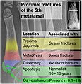
English: edit Proximal fractures of 5th metatarsal: Proximal fractures of the fifth metatarsal are common,[1] and are distinguished by their locations:
Normal anatomy that may simulate a fracture include mainly:
Template in WikipediaTo edit image template in Wikipedia, go to: en:Template:Image of proximal fractures of the 5th metatarsal. Further reading |
|||||
| Date | ||||||
| Source |
edit References
|
|||||
| Author |
 - Reusing images - Conflicts of interest: None |
|||||
| Other versions |
|
Licensing
| This file is made available under the Creative Commons CC0 1.0 Universal Public Domain Dedication. | |
| The person who associated a work with this deed has dedicated the work to the public domain by waiving all of their rights to the work worldwide under copyright law, including all related and neighboring rights, to the extent allowed by law. You can copy, modify, distribute and perform the work, even for commercial purposes, all without asking permission.
http://creativecommons.org/publicdomain/zero/1.0/deed.enCC0Creative Commons Zero, Public Domain Dedicationfalsefalse |
Captions
Items portrayed in this file
depicts
28 July 2019
File history
Click on a date/time to view the file as it appeared at that time.
| Date/Time | Thumbnail | Dimensions | User | Comment | |
|---|---|---|---|---|---|
| current | 08:06, 29 July 2019 |  | 1,709 × 1,772 (666 KB) | Mikael Häggström | Singular |
| 08:03, 29 July 2019 |  | 1,709 × 1,772 (674 KB) | Mikael Häggström | User created page with UploadWizard |
File usage
The following 4 pages use this file:
Global file usage
The following other wikis use this file:
- Usage on az.wikipedia.org
Metadata
This file contains additional information, probably added from the digital camera or scanner used to create or digitize it.
If the file has been modified from its original state, some details may not fully reflect the modified file.
| Software used | www.inkscape.org |
|---|


