Jump to content

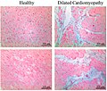
File:Histopathology of interstitial fibrosis in dilated cardiomyopathy.jpg

Page contents not supported in other languages.
This is a file from the Wikimedia Commons
From Wikipedia, the free encyclopedia

Original file (913 × 777 pixels, file size: 205 KB, MIME type: image/jpeg)

Summary

Description
English: Histopathology of interstitial fibrosis in dilated cardiomyopathy. Original caption:
Figure 1. Alcian blue staining showing areas of glycosaminoglycan (GAG) accumulation in hearts from patients with dilated cardiomyopathy (DCM) compared to healthy (non-failing control, NFC) hearts.
Date
Source (2019). "The Non-Fibrillar Side of Fibrosis: Contribution of the Basement Membrane, Proteoglycans, and Glycoproteins to Myocardial Fibrosis". Journal of Cardiovascular Development and Disease 6 (4): 35. DOI:10.3390/jcdd6040035. ISSN 2308-3425.
- Attribution 4.0 International (CC BY 4.0)
Author Michael Chute, Preetinder Aujla, Sayantan Jana and Zamaneh Kassiri

Licensing

w:en:Creative Commons
attribution
This file is licensed under the Creative Commons Attribution 4.0 International license.
You are free:
  • to share – to copy, distribute and transmit the work
  • to remix – to adapt the work
Under the following conditions:
  • attribution – You must give appropriate credit, provide a link to the license, and indicate if changes were made. You may do so in any reasonable manner, but not in any way that suggests the licensor endorses you or your use.

Captions

Histopathology of interstitial fibrosis in dilated cardiomyopathy

Items portrayed in this file

depicts

23 September 2019

image/jpeg

File history

Click on a date/time to view the file as it appeared at that time.

Date/TimeThumbnailDimensionsUserComment
current08:01, 31 January 2020Thumbnail for version as of 08:01, 31 January 2020913 × 777 (205 KB)Mikael HäggströmUser created page with UploadWizard

The following 3 pages use this file:

Global file usage

The following other wikis use this file: