Jump to content

File:F29-03-9780123846846-Rudiviridae-Fig3-SIRV2-infection.png

Page contents not supported in other languages.
This is a file from the Wikimedia Commons
From Wikipedia, the free encyclopedia

Original file (1,975 × 1,936 pixels, file size: 880 KB, MIME type: image/png)

Summary

Description
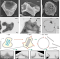
English: Transformation of Sulfolobus host cells following infection with Sulfolobus islandicus rod-shaped virus 2 (SIRV2, Rudiviridae): (A1, A2), uninfected cells; (B1, B2), cells 10 h post-infection; (C1,C2), cells 10–14 h post-infection. (A1, B1, C1) scanning electron micrographs; (A2, B2, C2) transmission electron micrographs of thin sections of negatively stained cells. Scale bars: 200 nm. The electron micrographs are accompanied by the schematic representation of the cell at different stages of the virus replication cycle. Times after infection are indicated in hours. At 0 h, two chromosomes of the Sulfolobus host are shown in blue. Later they degrade concomitantly with formation of virus-induced pyramids (shown in red) and the intracellular clusters of assembled virions. Finally, at time points between 10 and 14 h, the virus-induced pyramids open (their remains are shown in red), the cell lyses and the virions are extruded. The gradual opening out of the virus-induced pyramids (at time points between 10 and 14 h) is illustrated in more details with fragments from electron micrographs of thin sections of the infected cell.
Date
Source https://talk.ictvonline.org/cfs-file/__key/communityserver-wikis-components-files/00-00-00-00-21/f29_2D00_03_2D00_9780123846846.png at https://talk.ictvonline.org/ictv-reports/ictv_9th_report/dsdna-viruses-2011/w/dsdna_viruses/132/rudiviridae-figures ICTV 9th Report (2011) Rudiviridae - Figures: Fig. 3
Author International Committee on Taxonomy of Viruses (ICTV): Modified from Vestergaard et al. (2008). J. Bacteriol., 190, 6837-6845.
Other versions


Licensing

w:en:Creative Commons
attribution share alike
This file is licensed under the Creative Commons Attribution-Share Alike 4.0 International license.
You are free:
  • to share – to copy, distribute and transmit the work
  • to remix – to adapt the work
Under the following conditions:
  • attribution – You must give appropriate credit, provide a link to the license, and indicate if changes were made. You may do so in any reasonable manner, but not in any way that suggests the licensor endorses you or your use.
  • share alike – If you remix, transform, or build upon the material, you must distribute your contributions under the same or compatible license as the original.

Captions

Add a one-line explanation of what this file represents

Items portrayed in this file

depicts

File history

Click on a date/time to view the file as it appeared at that time.

Date/TimeThumbnailDimensionsUserComment
current10:51, 24 March 2021Thumbnail for version as of 10:51, 24 March 20211,975 × 1,936 (880 KB)ErnstsUploaded a work by International Committee on Taxonomy of Viruses (ICTV): Modified from Vestergaard ''et al.'' (2008). J. Bacteriol., 190, 6837-6845. from https://talk.ictvonline.org/cfs-file/__key/communityserver-wikis-components-files/00-00-00-00-21/f29_2D00_03_2D00_9780123846846.png at https://talk.ictvonline.org/ictv-reports/ictv_9th_report/dsdna-viruses-2011/w/dsdna_viruses/132/rudiviridae-figures ICTV 9th Report (2011) Rudiviridae - Figures: Fig. 3 with UploadWizard

The following page uses this file:

Global file usage

The following other wikis use this file:

Metadata