File:Diaphragm Arthur Keith 1.jpg
Diaphragm_Arthur_Keith_1.jpg (559 × 325 pixels, file size: 32 KB, MIME type: image/jpeg)
| Description |
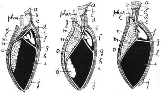
Figs. 1, 2, 3 -- Diagrams illustrating the origin of the diaphragm and pleural cavities. Fig. 1, the amphibian body-cavities; fig. 2, the corresponding cavities in a bird; fig. 3, in a mammal. a, mandible; b, genio-hyoid; c, hyoid; d, sterno-hyoid; e, sternum; f, pericardium; g, septum transversum; h, rectus abdominalis; i, abdominal cavity; j, pubis; k, œsophagus; l, trachea; m, cervical limiting membrane of abdominal cavity; n, dorsal wall of body; o, lung; o', air-sac. | ||
|---|---|---|---|
| Source |
From "The nature of the mammalian diaphragm and pleural cavities", Arthur Keith, M.D., Journal of Anatomy and Physiology, 1905.[1] | ||
| Date | |||
| Author |
| ||
| Permission (Reusing this file) |
See below.
|
| This image is in the public domain in the United States because it was first published outside the United States prior to January 1, 1929. Other jurisdictions have other rules. Also note that this image may not be in the public domain in the 9th Circuit if it was first published on or after July 1, 1909 in noncompliance with US formalities, unless the author is known to have died in 1953 or earlier (more than 70 years ago) or the work was created in 1903 or earlier (more than 120 years ago.)[2] |
File history
Click on a date/time to view the file as it appeared at that time.
| Date/Time | Thumbnail | Dimensions | User | Comment | |
|---|---|---|---|---|---|
| current | 15:22, 22 October 2007 |  | 559 × 325 (32 KB) | Mike Serfas (talk | contribs) | {{PD-US-1923-abroad}} The nature of the mammalian diaphragm and pleural cavities, Arthur Keith, M.D., Journal of Anatomy and Physiology, 1905.[http://books.google.com/books?id=2ecDAAAAYAAJ] Legend (not included in this image): Figs. 1, 2, 3 -- Diagrams |
| 15:19, 22 October 2007 |  | 559 × 485 (53 KB) | Mike Serfas (talk | contribs) | {{US-PD-1923-abroad}} The nature of the mammalian diaphragm and pleural cavities, Arthur Keith, M.D., Journal of Anatomy and Physiology, 1905.[http://books.google.com/books?id=2ecDAAAAYAAJ] Legend (not included in this image): Figs. 1, 2, 3 -- Diagrams |
You cannot overwrite this file.
File usage
The following page uses this file:


