Jump to content

File:Bathyhedyle boucheti (10.7717-peerj.2738) Figure 2.png

Page contents not supported in other languages.
This is a file from the Wikimedia Commons
From Wikipedia, the free encyclopedia

Original file (1,963 × 2,623 pixels, file size: 4.01 MB, MIME type: image/png)

Summary

Description
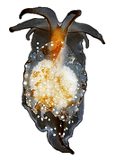
English: Figure 2: Photograph of a living specimen (A) and 3D reconstructions (B–G) of Bathyhedyle boucheti n. sp.
(A) External morphology, dorsal view. (B) General microanatomy, dorsal view, (C) right view. (D–G) Positions of the organ systems, dorsal view; (D) central nervous system, (E) digestive system, (F) circulatory and excretory systems, (G) reproductive system. a, anus; dv, ‘dorsal vessel system’; ey, eye; f, foot; fgo, female gonopore; gl, subepidermal gland; h, heart; lt, labial tentacle; mgo, male gonopore; n, notum; ov, ovotestis; p, penis; pn, pedal nerve; pt, propodial tentacle; rh, rhinophore; rhn, rhinophoral nerve. Scale bars: (A–D) 1 mm, scale bar in (D) valid for (D–G).
Date
Source https://doi.org/10.7717/peerj.2738
Author Neusser TP, Jörger KM, Lodde-Bensch E, Strong EE, Schrödl M. 2016. The unique deep sea—land connection: interactive 3D visualization and molecular phylogeny of Bathyhedyle boucheti n. sp. (Bathyhedylidae n. fam.)—the first panpulmonate slug from bathyal zones. PeerJ 4:e2738 https://doi.org/10.7717/peerj.2738
Permission
(Reusing this file)
w:en:Creative Commons
attribution
This file is licensed under the Creative Commons Attribution 4.0 International license.
You are free:
  • to share – to copy, distribute and transmit the work
  • to remix – to adapt the work
Under the following conditions:
  • attribution – You must give appropriate credit, provide a link to the license, and indicate if changes were made. You may do so in any reasonable manner, but not in any way that suggests the licensor endorses you or your use.
Other versions

Captions

Add a one-line explanation of what this file represents

Items portrayed in this file

depicts

6 December 2016

image/png

14fcbbc5242299fce3900dcf2e892fbbaf333dc6

4,201,924 byte

2,623 pixel

1,963 pixel

File history

Click on a date/time to view the file as it appeared at that time.

Date/TimeThumbnailDimensionsUserComment
current14:28, 13 November 2020Thumbnail for version as of 14:28, 13 November 20201,963 × 2,623 (4.01 MB)Christian Ferrer{{Information | description = {{en|1= Figure 2: Photograph of a living specimen (A) and 3D reconstructions (B–G) of ''Bathyhedyle boucheti'' n. sp. :(A) External morphology, dorsal view. (B) General microanatomy, dorsal view, (C) right view. (D–G) Positions of the organ systems, dorsal view; (D) central nervous system, (E) digestive system, (F) circulatory and excretory systems, (G) reproductive system. a, anus; dv, ‘dorsal vessel system’; ey, eye; f, foot; fgo, female gonopore; gl, subepide...
No pages on the English Wikipedia use this file (pages on other projects are not listed).